Heparin is a pharmaceutical drug that is used to prevent the formation of blood clots [1]. Since diseases that can affect the circulatory system are widespread nowadays, it is important to effectively monitor blood coagulation, especially during surgery. Methods that are currently used for heparin detection have a few drawbacks. Firstly, these methods are not specific for heparin and there are loads of analytical interferences. Secondly, they are time consuming [2]. These drawbacks led to a need for a new analytical system that is specific for heparin. Since it is a highly negatively charged polysaccharide, heparin can be easily adapted to the analytical system where negatively charged colloidal gold nanoparticle (GNP) solution is used resulting in aggregation [3].
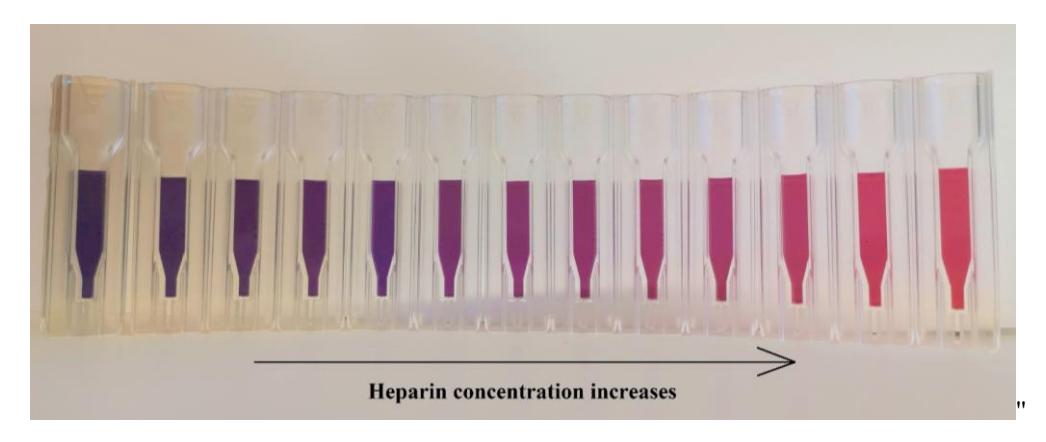
In this work, we compared how the difference in GNPs size (13 nm and 3.5 nm) affect analytical parameters such as sensitivity and reproducibility of the system used for heparin detection. The system itself relies on the coagulation of negatively charged colloidal GNP solution in the presence of positively charged polymer poly-L-lysine (PLL). Stable GNP solution is red due to the local surface plasmon resonance (LSPR) and the peak is registered at a wavelength of 520 nm. The addition of PLL to GNP solution results in nanoparticle aggregation. Visible color change can be observed after the aggregation with GNP solution turning blue and LSPR peak shifting to a wavelength of 650 nm. On the other hand, heparin stabilizes GNP solution by preventing the PPL induced aggregation, resulting in a GNP solution of specific color based on heparin concentration allowing for the creation of a heparin sensor (Fig. 1).