Viscosity imaging at a microscopic scale can reveal information about the diffusion-controlled processes in biosystems. Determining a microviscosity change can indicate the development of atherosclerosis, diabetes, and Alzheimer's disease.[1] 'Molecular rotors' are viscosity-sensitive fluorophores that provide one of the easiest methods to image microviscosity. These fluorophores have been employed for imaging microviscosity in polymers, living cells and lipid bilayers.[1] The concept of molecular rotors mechanism is based on a changing fluorescence signal, which emerges from the competition between intramolecular rotation and emitted photons. The nature of the electronically excited state is changed by the rotation causing a faster non-radiative relaxation. Therefore, a longer fluorescence lifetime of the molecule is observed in a high viscosity environment.[2]
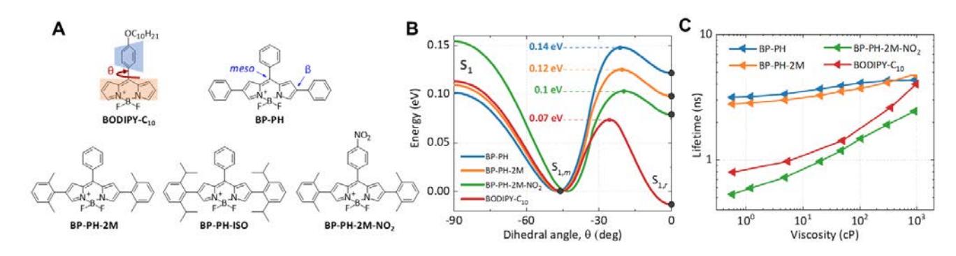
One of the most widely-used molecular rotors is BODIPY-C10 (Fig. 1A). This standout molecule displays the monoexponential fluorescence decay, which simplifies the data analysis. Unfortunately, the green absorbance and fluorescence wavelengths are the greatest drawbacks of the BODIPY-C10. It is well known, that the longer wavelengths are desired for viscosity imaging of the biological sample. It is possible to achieve longer wavelengths by introducing conjugated substituents and extending the conjugation length within the fluorophore.[3] However, we have to ensure that the new red-emitting BODIPY molecule remains viscosity-sensitive.
To avoid a time-consuming synthesis of a large number of fluorophores with various modifications, we show how density functional theory (DFT) calculations can help to determine viscosity-sensitive properties of the molecule before its synthesis. In this work, we investigate four phenyl-substituted BODIPY molecular rotors (Fig. 1A): without any additional moieties on β-phenyls (BP-PH), with two methyl (BP-PH-2M) or isopropyl (BP-PH-ISO) groups on each β-phenyl ring, and methyl-substituted β-phenyls with a nitro-substituted meso-phenyl (BP-PH-2M-NO2). In this case, adding β-phenyls increases molecule's conjugation and red-shifts fluorescence spectra to biologically-friendly wavelengths.
The research consists of quantum chemical calculations, absorption and fluorescence spectra measurements, fluorescence lifetime evaluation, and live cells imaging using lipid vesicles. Dependencies regarding the molecular structure, activation energy barrier, increasing solvent viscosity, polarity, and temperature were investigated. Quantum chemical calculations showed that an increase in an activation energy barrier (Fig. 1B) results in red-shifted fluorescence spectra. Moreover, DFT-based calculations allowed us to create a red-emitting probe by introducing a nitro group, which reduces the barrier and increases viscosity-sensitivity of the molecule (Fig. 1C).[4]