ELECTRON PARAMAGNETIC RESONANCE: GOING MICRO FOR MACRO GAINS
Gediminas Usevičius1, Oscar W. Kennedy2, Ignas Pocius1, Patrick Hogan2, Ana Villanueva Ruiz de Temino2, Jean-Baptiste Verstraete2, Paulina Verbaitytė1, G. Antilen Jacob2, Mindaugas Kamarauskas3, Marius Treideris3, Joseph Alexander2, Vidmantas Kalendra1, Jūras Banys1, John J.L. Morton2, 4, Mantas Šimėnas1
1 Faculty of Physics, Vilnius University, Sauletekio 3, LT-10257 Vilnius, Lithuania
2 London Centre for Nanotechnology, University College London, London WC1H 0AH, UK
3 Center for Physical Sciences and Technology, Sauletekio 3, LT-10257 Vilnius, Lithuania
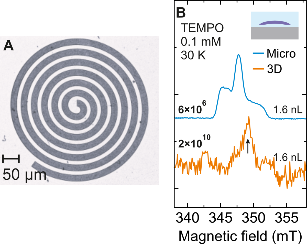
The increasing demand of quantum technologies and a need to study minute amount samples has not surpassed electron paramagnetic resonance (EPR) spectroscopy [1]. 3D resonators with few \(\mu\)L mode volumes rarely do the job when studying only tens of billion spins. Thus, in recent years microresonators of various geomteries have been desgined and used to study systems previously unfeasible to do so [2,3]. However, their application remained very limited due to requiring very specific instrumental setups and samples. Here, I present our approach which employs planar spiral-shaped microresonators of 7 nL mode volumes fabricated from Yttrium Barium Copper Oxide (YBCO) high-temperature superconductor. A significant 3500-fold increase in the spin number sensitivity over commercial 3D resonators has been achieved which is fully compatible with conventional instrumentation and typical sample conditions. Our approach significantly advances the applicability of superconducting microresonators as versatile and readily applicable tools for high sensitivity EPR.
Fig. 1. (A) Fabricated planar YBCO spiral microwave microresonator on a sapphire substrate.(B) Echo-detected field sweep spectra of a 0.1 mM TEMPO sample obtained at 30 K using the spiral microresonator (1.6 nL sample volume) (blue) and a measurement of the same sample obtained using the Bruker MD5 resonator and the same measurement time are presented (orange) for comparison yielding only the impurity signal of the sapphire ring (arrows). The determined spin-number sensitivities (spins/G/$\sqrt{\text{Hz}}$) are indicated above the measured spectra.
[1] D. Goldfarb and S. Stoll, EPR Spectroscopy: Fundamentals and Methods, eMagRes Books. Wiley, 2018.
[2] Y. Twig, E. Suhovoy, and A. Blank, “Sensitive surface loop-gap microresonators for electron spin resonance,” Rev. Sci. Instrum., vol. 81, p. 104703, 2010.
[3] A. Bienfait et al., “Reaching the quantum limit of sensitivity in electron spin resonance,” Nat. Nanotechnol., vol. 11, pp. 253–257, 2016.