
Due to an increasingly changing environment, plants are constantly exposed to adverse environmental conditions that can impair photosynthesis and lead to the overproduction of reactive oxygen species (ROS). This complex phenomenon is known as oxidative stress and can cause significant damage to cellular structures, including chloroplasts, thereby affecting plant growth and development [1]. Implementing early non-invasive detection techniques could assist in the development of innovative strategies to mitigate the effects of adverse environmental conditions and enhance plant resilience.
The effect of ROS was evaluated at the single cell level, employing internodal cells of green macroalga Nitellopsis obtusa. Microscopic images were taken to visualize the possible damage of externally applied H\(_{2}\)O\(_{2}\) (250 µM and 500 µM) on the chlorophyll layer of algal cells after 24 h incubation. In addition, changes in the concentrations of carotenoids and chlorophylls in cell extracts were evaluated via absorption spectroscopy. Photosynthetic processes of intact algal cells after 6 h incubation in H\(_{2}\)O\(_{2}\) were monitored via pulse-amplitude-modulated (PAM) fluorometry, using blue LED light (445 nm) for excitation.
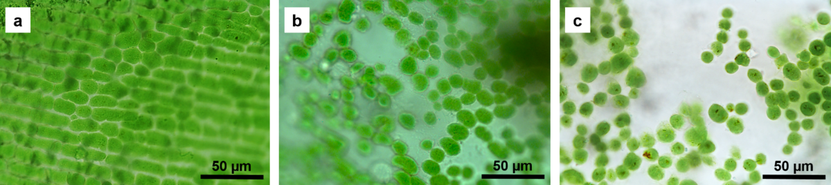
Microscopic images showed that H\(_{2}\)O\(_{2}\) damaged the chloroplast layers of algal cells after prolonged exposure (24 h) (Fig. 1). Spectroscopic analysis revealed that H\(_{2}\)O\(_{2}\) at a concentration of 500 µM caused a reduction in chlorophyll content over 24 h, while both applied concentrations (250 µM and 500 µM) resulted in a significant decrease in carotenoid concentration (p < 0.05, Kruskal–Wallis test). Fluorometry showed that the quantum yield of PSII (Y(II)), a measure of photosynthetic efficiency, was lower in both groups studied compared to the values registered in control cells (p < 0.05, Kruskal–Wallis test).
The study conducted shows that H\(_{2}\)O\(_{2}\) (250 µM and 500 µM) causes oxidative stress in algal cells, as indicated by a decrease in carotenoid concentrations, which have antioxidant properties [2]. The effects studied were detected in viable cells using non-invasive techniques, suggesting that the applied methods may be applicable for early detection of physiological stress in plants or for detection of lower, environmentally relevant concentrations of ROS.