Supramolecular organization of π-conjugated molecules is an attractive way to create functional materials by amplifying the effects of the inherent molecular functionality. Among various π-conjugated molecules TPPS are especially interesting because they have characteristic bands in the visible region, making their supramolecular polymers fascinating candidates for practical electro-optical materials. Over the years, many porphyrin-based supramolecular materials have been created by tuning structures of their building blocks (monomers), especially by varying the peripheral groups attached to the porphyrin core. The number of peripheral SO\(^{-}_{3}\) groups influences properties of TPPS molecules that can change their practical applications [1]. Here we report differences in protonation process of the porphyrin centre that is affected by number of SO\(^{-}_{3}\) groups in TPPS molecules– 5,10,15,20-tetrakis(4-sulfonatophenyl)porphyrin (TPPS\(_{4}\)), 5,10,15-tris(4-sulfonatophenyl)-20-phenylporphyrin (TPPS\(_{3}\)) and 5,10-bis(4-sulfonatophenyl)-15,20-diphenylporphyrin (TPPS\(_{2a}\)).
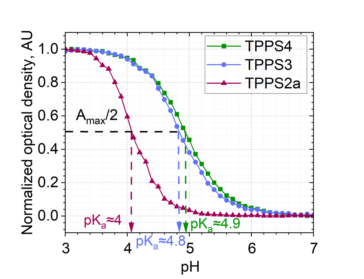
In this study absorbance spectra of TPPS\(_{4}\), TPPS\(_{3}\), TPPS\(_{2a}\) was continuously measured during titration with hydrochloric acid and sodium hydroxide. Bi-directional titration experiments were performed: from pH=3 to pH=7 (using NaOH) and from pH=7 to pH=3 (using HCl). Changes of absorbance intensity of 434 nm Soret band upon titration was used for determination of pK\(_{a}\) values. Upon acidic titration Soret band shifts from 414 nm (not protonated form) to 434 nm (protonated form). Middle point of this change indicates pK\(_{a}\) value– pH at which concentrations of free base and protonated forms are identical. We determined following pK\(_{a}\) values: pK\(_{a}\)=4.9 for TPPS\(_{4}\), pK\(_{a}\)=4.8 for TPPS\(_{3}\) and pK\(_{a}\)=4 for TPPS\(_{2a}\). This difference of pK\(_{a}\) is likely caused by different net charge and different charge distribution of TPPS\(_{4}\), TPPS\(_{3}\) and TPPS\(_{2a}\). TPPS\(_{4}\) acquires biggest net charge (-4) which is created by four SO\(^{-}_{3}\) groups. Bigger negative net charge at the periphery of molecule might cause electron density redistribution and lead to more favourable conditions for protonation of the porphin core.
The acquired results indicate that higher number of peripheral SO\(^{-}_{3}\) groups favours protonation of the core of TPPS at higher pH value, at lower H\(^{+}\) concentrations. This also leads to formation of supramolecular TPPS assemblies at higher pH values via ionic interaction between protonated porphyrin core with negatively charged SO\(^{-}_{3}\).