
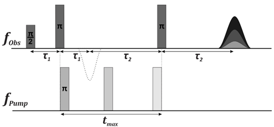
Double electron-electron resonance (DEER) spectroscopy is a powerful and widely used method in structural biology, providing insights into the structural and dynamic properties of biomolecules such as proteins, RNA, and DNA [1]. This pulsed electron paramagnetic resonance (EPR) technique enables the measurement of nanoscale distances between unpaired electron spins by employing microwave pulses at different frequencies. One electron spin is detected while the other is excited, allowing for the observation of dipolar interactions throughout the experiment (Fig. 1) [2]. Since many biomolecules lack unpaired electrons, DEER spectroscopy utilizes site-directed spin labeling (SDSL) to introduce spin labels, enabling structural investigations that are not constrained by crystallization requirements or molecular weight limitations [1].
In this study, DEER spectroscopy was used to examine the calcium-binding proteins S100A8 and S100A9, which are associated with neurodegenerative disorders such as Alzheimer’s and Parkinson’s diseases. Using nitroxide radical spin labels, we measured the distance distributions between labeled cysteine residues, revealing key structural and dynamic properties of these proteins. Our findings demonstrate the potential of DEER spectroscopy for studying protein conformational changes, interactions, and functional mechanisms, further contributing to the understanding of their role in disease-related processes.