The intracellular environment’s physical properties, particularly microviscosity, regulate molecular diffusion and cell function. Microviscosity varies across organelles, changes under pathological conditions, and shifts during mesenchymal stem cell (MSC) differentiation [1]. MSCs are highly plastic, adapting their shape to biomechanical cues, which influence differentiation, proliferation, and organisation of intracellular structures. However, it remains unknown how MSC shape affects microviscosity in different organelles.
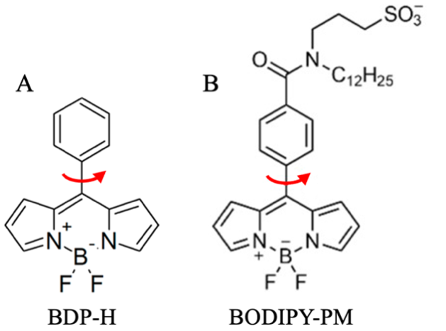
In this study, we used fluorescence lifetime imaging microscopy (FLIM) with two viscosity-sensitive fluorophores, BDP-H (Fig. 1A) and BODIPY-PM (Fig. 1B), to measure microviscosity in MSCs. Both BDP-H and BODIPY-PM change their fluorescence lifetime in response to microviscosity changes, displaying biexponential fluorescence decays in lipid droplets and plasma membrane, respectively [2-3].
The aim of our research was to measure the microviscosity of lipid droplets and plasma membranes in human skin-derived MSCs cultured on adhesive micropatterns of varying shapes and sizes.
MSCs were grown on polyethylenglycol hydrogel with covalently immobilized adhesion promoting fibronectin micropatterns shaped like circles, drops and triangles, each available in three sizes: 710 μm\(^2\), 1260 μm\(^2\), 2000 μm\(^2\). Live-cell imaging was conducted using a laser-scanning confocal microscope, and fluorescence lifetimes were measured using time-correlated single photon-counting based FLIM.
Our results show that the fluorescence lifetime of BDP-H remained unchanged across different micropatterns, indicating that lipid droplet microviscosity is unaffected by cell shape or size. In contrast, BODIPY-PM fluorescence lifetime varied significantly with cell geometry and size, demonstrating that plasma membrane microviscosity is highly dynamic and responsive to morphological changes. Taken together, our findings suggest that organelles respond differently to morphological changes, with manipulation of cell geometry through adhesive micropatterns specifically altering plasma membrane microviscosity but not lipid droplet microviscosity.