Differentiation of cells completely alters their morphological and biochemical profiles. While the aforementioned profiles have been sufficiently characterised, the biomechanical remodelling, such as the change of viscosity within cell structure and organelles, remains unmonitored. Determining viscosity during and after differentiation may provide deeper insight into the vital process, especially for stem cells, e.g. mesenchymal stem cells (MSC) that are capable of differentiating into fat, bone, cartilage or other connective tissue. Therefore, a state-of-the-art method is essential for a spatial resolved quantitative intracellular viscosity 'mapping' in MSC and their differentiated counterparts.
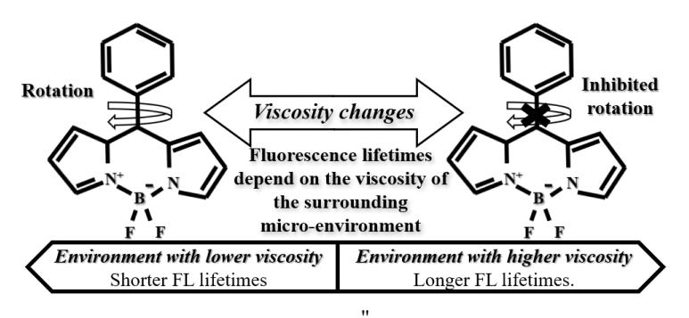
Molecular rotors are viscosity-sensitive fluorophores that may be utilized in monitoring intracellular viscosity. One of such molecules - BODIPY-h (BDP-h) is based on BODIPY (4,4-difluoro-4-bora-3a,4a-diaza-s-indacene) fluorescent dye. The molecule was tested in methanol-glycerol and toluene-castor oil mixtures and its sensitivity to viscosity was confirmed [1]. BDP-h phenyl ring's rotation depends on the viscosity of the surrounding micro-environment (Fig. 1): more viscous medium inhibits intramolecular rotation, which leads to slower deactivation from the fluorescent state via non-radiative energy transition, thus resulting in a longer fluorescence (FL) lifetime and vice versa [2].
The aim of our study was to determine the photophysical properties of BDP-h in aqueous media, such as phosphate-buffered saline (PBS) or cell growth medium - Dulbecco's Modified Eagle Medium (DMEM) with or without fetal bovine serum (FBS), together with the uptake of the molecular rotor in human skin MSC and their differentiated counterparts: adipocytes, osteocytes and chondrocytes.
Absorption, fluorescence spectra and fluorescence lifetimes of BDP-h were measured in distilled water, PBS and DMEM with or without FBS. For the uptake evaluation, StemPro differentiation kits (Gibco, US), were applied for specific differentiation of MSC. Cells were stained with 9 µM BODIPY-h solution diluted with DMEM (1:1000) (Gibco, US) and incubated for 120 min. The accumulation of dye was observed using Nikon Eclipse Te2000-S confocal microscope (Nikon, Japan).
Our findings indicate that molecular rotor BDP-h interacts with serum proteins, which results in a slight red shift of both BDP-h absorption and fluorescence spectra by 6 nm and 4 nm, respectively. Inclusion of protein molecules also results in longer fluorescence lifetimes, which increases from 0.22 ns to 4.7 ns. BDP-h uptake evaluation revealed that dye accumulates both in fixed and live MSC. The molecular rotor diffuses through the membrane and stains membrane-bound organelles without passing the nuclear membrane. In differentiated MSC the intracellular distribution is different: in adipocytes BDP-h accumulates in lipid droplets, while in chondrocytes and osteocytes the dye exhibits cytosolic staining.