THIRD-HARMONIC GENERATION MICROSCOPY OF COLLAGEN IN BIOLOGICAL TISSUES STAINED WITH HEMATOXYLIN AND EOSIN
Mykolas Mačiulis1, Viktoras Mažeika1, Martynas Riauka1, Lukas Kontenis1, 2, Danutė Bulotienė1, 3, Vitalijus Karabanovas1, 3, 4, Edvardas Žurauskas5, Virginijus Barzda1, 6, 7
1 Laser Research Centre, Faculty of Physics, Vilnius University, Saulėtekio Ave. 10, LT-10223, Vilnius, Lithuania
2 Light Conversion, 2B Keramikų st., LT-10233, Vilnius, Lithuania
3 Biomedical Physics Laboratory, National Cancer Institute, P. Baublio st. 3b, LT-08406, Vilnius, Lithuania
4 Department of Chemistry and Bioengineering, Vilnius Gediminas Technical University, Saulėtekio Ave. 11, LT-10223, Vilnius, Lithuania
5 Department of Pathology, Forensic Medicine and Pharmacology, Faculty of Medicine, Vilnius University, M.K. Čiurlionio st. 21/27, LT-03101, Vilnius, Lithuania
6 Department of Physics, University of Toronto, 60 St. Georges st., Toronto, ON M5S 1A7, Canada
7 Department of Chemical and Physical Sciences, University of Toronto Mississauga, 3359 Mississauga Rd. N., Mississauga L5L 1C6, ON, Canada
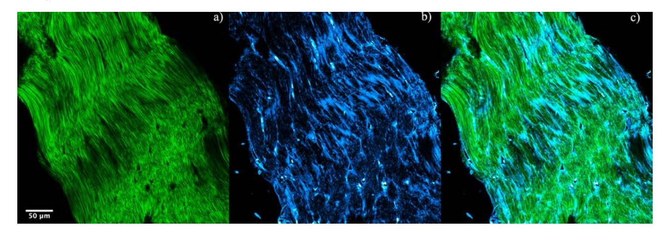
Multimodal nonlinear laser-scanning microscopy is a powerful technique for noninvasive biological imaging [1]. Several nonlinear signals can be generated simultaneously at the focus of a microscope objective, including multiphoton excitation fluorescence, second-harmonic generation (SHG) and third-harmonic generation (THG). Collagen, the main constituent of extracellular matrix in the biological tissue can be visualized with SHG microscopy, which can be used for cancer diagnostics [2]. Changes in the collagen structure are observed in hematoxylin-and-eosin-stained (H&E-stained) histology sections, which contrast cell nuclei from the cytoplasm and extracellular structures and are considered as the gold standard for histological investigations [3]. Although collagen fibers are visualized with SHG microscopy, in this work THG is used for collagen visualization and characterization. The potential for cancer diagnostics using THG from H&E-stained samples is also investigated. Fig.1(a) visualizes collagen fibrils in SHG, while THG (Fig.1(b)) shows endotendineum which wraps individual fascicles. Collagen fibrils are also faintly visible in the THG image. In addition cell nuclei of tendocytes are highlighted in THG. The combined image (Fig.1(c)) shows the colocalization between collagen fibrils, endotendineum and tendocytes. Multimodal nonlinear microscopy imaging can be incorporated into the tissue histology investigations without additional modifications of the H&E-stained histopathology samples. Multimodal imaging can be applied for investigation of structural changes in connective tissue due to various diseases and also is beneficial in observing extracellular matrix and cell nuclei in cancer diagnostics.
Fig. 1. Multicontrast nonlinear optical microscopy images of rat tail tendon histology section stained with hematoxylin and eosin (H&E staining). a) SHG image of collagen tissue, b) THG image from the same sample area and c) combined image of both.
Acknowledgments: The work was supported by grant No. 1.2.2.-LMT-K-718-02-0016.
[1] R. Carriles, D. N. Schafer, K. E. Sheetz, J. J. Field, R. Cisek, V. Barzda, and J. Squier, "Imaging techniques for harmonic and multi-photon absorption fluorescence microscopy," Rev. Sci. Instrum.80, 081101 (2009).
[2] A. Golaraei, L. Kontenis, R. Cisek, D. Tokarz, S. J. Done, B. C. Wilson, and V. Barzda, "Changes of collagen ultrastructure in breast cancer tissue determined by second-harmonic generation double Stokes-Mueller polarimetric microscopy," Biomed. Opt. Express7(10), 4054-4068 (2016).
[3] J. A. Kiernan, Histological and Histochemical Methods, 3rd ed., Butterworth Heinemann, Woburn, MA (1999).