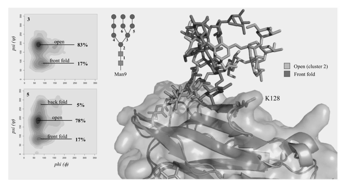
Oligomannoses are evolutionarily the oldest class of N-glycans, where the arms of the common pentasaccharide unit, i.e. Manα(1-6)-[Manα(1-3)]-Manβ(1-4)-GlcNAcβ(1-4)-GlcNAcβ1-Asn, are functionalized exclusively with branched arrangements of mannose (Man) monosaccharide units. In mammalian species oligomannose N-glycans can have up to 9 Man, meanwhile structures can grow to over 200 units in yeast mannan. The highly dynamic nature, branching complexity and 3D structure of oligomannoses have been recently highlighted for their roles in immune escape and infectivity of enveloped viruses, such as SARS-CoV2 and HIV-1. The architectural features that allow these N-glycans to perform their functions is yet unclear, due to their intrinsically disordered nature that hinders their structural characterization. In this work we will discuss the results of over 54 μs of cumulative sampling by molecular dynamics (MD) simulations of differently processed, unlinked oligomannose N-glycans common in vertebrates. We then discuss the effects of a complex protein surface on their structural equilibria based on over 4 μs cumulative MD sampling of the fully glycosylated CD16a Fc gamma receptor (FcγRIIIa), where the type of glycosylation is known to modulate its binding affinity for IgG1s, regulating the antibody-dependent cellular cytotoxicity (ADCC). Our results show that the protein's structural constraints shift the oligomannoses conformational ensemble to promote conformers that satisfy the steric requirements and hydrogen bonding networks demanded by the protein's surface landscape. More importantly, we find that the protein does not actively distort the N-glycans into structures not populated in the unlinked forms in solution. Ultimately, the highly populated conformations of the Man5 linked glycans support experimental evidence of high levels of hybrid complex forms at N45 and show a specific presentation of the arms at N162, which may be involved in mediating binding affinity to the IgG1 Fc.