Zinc oxide is an important semiconducting material which is widely used in catalysis, photovoltaics, and pigment industry [1]. The application of ZnO nanoparticles depends on their structure and morphology, which can be varied by changing film formation methods.
The aim of this work is to compare properties of ZnO coatings deposited on AISI 304 stainless steel by electrochemical deposition (ECD) and electrophoretic deposition (EPD). Electrochemical measurements were performed by a computer controlled Autolab PGSTAT12 (Ecochemie, The Netherlands) potentiostat/galvanostat using a standard three electrode cell (volume 100 mL). All the potentials are referred to the Ag, AgCl KCl (sat) reference electrode. The surface morphology and the composition of ZnO powder were investigated with a Quanta FEG 200 (FEI) high resolution scanning electron microscope (SEM) that was equipped with a Bruker XFlash® 4030 detector (Bruker AXS) for high resolution energy dispersive X-ray spectroscopy (EDX). X-ray powder diffraction (XRD) data were collected with a DRON-6 (Bourevestnik Inc., Russia) powder diffractometer equipped with Bragg-Brentano geometry and using Ni-filtered CuKα radiation and a graphite monochromator. The photoelectrochemical activity of the prepared electrodes was investigated by employing photovoltammetry measurement methods. A photoelectrochemical quartz cell was employed. A General Electric F8W/BLB lamp (max=366 nm, power density 1.8 mW·cm-2) was placed at a distance of 2 cm from the ZnO electrode and was used as an UV radiation source [2].
Electrochemically deposited zinc oxide coatings were produced using slightly acidic zinc (II) acetate solution under galvanostatic conditions - the current density was 1.0 mA·cm-2 and duration time - 10 min. Homogeneous suspension (2 g of the ZnO powder dispersed in 100 mL of methanol) was used for the electrophoretic deposition of ZnO coatings and the potential of 30 V was applied for 20 minutes. In order to achieve better adhesion of ZnO particles on stainless steel deposited by ECD and EPD were annealed at 400 °C for 1 h in the atmospheric air.
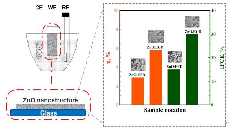
XRD pattern of ECD and EPD ZnO films showed diffraction peaks at 2θ equal to 31.78°, 34.44° and 36.24°, which correspond to the well-crystallized wurtzite type ZnO (PDF 04-004-4120). However, the surface of ZnO coatings prepared by ECD and EPD differs. SEM images revealed that ECD ZnO film surface is granular composed of grains with diameter of 30-110 nm, whereas films prepared by EPD is flower-like structure mainly composed of rods.
Photoelectrochemical behavior of the ZnO electrode was determined from the current-potential curves obtained in 0.1 M Na2SO4 solutions both in the dark and under UV irradiation. Significant anodic current increase under UV irradiation shows n-type semiconducting properties. The calculated values of incident photon-to-current efficiency (IPCE) and applied bias photon-to-current conversion efficiency (${\eta}$) for the 0.6 V potential are shown in Figure 1. Electrochemically deposited ZnO films on stainless steel showed the best performance as a photoelectrode in 0.1 M Na2SO4 electrolyte.