
Second-harmonic generation (SHG) microscopy is a nonlinear imaging technique based on the frequency-doubling of photons interacting with non-centrosymmetric structures. It enables label-free in vivo and in situ visualization of non-centrosymmetric biopolymers such as collagen in most structural tissues and myosin filaments in the sarcomeres of striated muscles. Structural label-free nonlinear imaging emerges as a successful tool for faster biomedical diagnostics, while quantitative polarimetric imaging opens up new ways for more detailed tissue characterization.
The SHG process requires hight intensity coherent light. For this reason, imaging is commonly performed by raster-scanning a tightly focused femtosecond oscillator beam and acquiring the image pixel-by-pixel using a single-element photodetector. The frame rate is thus limited by the laser scanning speed. Increasing availability of robust, affordable, high-power, femtosecond lasers operating at MHz rates has stimulated the advancement of widefield SHG microscopy. Widefield microscopy does not require scanning and therefore enables real-time imaging of large samples [1]. A high frame rate is achievable because a large imaging area is exposed to short high intensity laser pulses and the image is obtained as a single frame using a camera. It should be noted that laser parameters need to be carefully optimised because high intensity laser radiation eventually damages the sample. The signal level can also be increased by using a higher repetition rate, leading to higher average power, which eventually causes photobleaching [2].
The purpose of this work was to develop a widefield microscope and to demonstrate its performance by imaging label-free biological samples. A PHAROS high-repetition-rate amplified laser system was used for evaluation of the developed widefield microscope by imaging label-free rat skeletal muscle tissue (Biomax RAT901a). The laser parameters at the sample were: 1 W average power, 100 kHz repetition rate, 680 μm beam diameter (1/e2), energy density of 5.5 μJ/cm2 and a peak intensity of 19 GW/cm2 using 290 fs pulses. We were able to produce 410 μm × 485 μm images with sub-micron resolution and with no observable damage to the sample. The image in figure 1(a,b) exhibits a periodic structure of the sarcomere anisotropic A-bands, which are clearly distinguishable from the dark isotropic I-bands due to SHG signal from myosin nanomotors. Figure 1(c) shows the SHG intensity profile of 20 sarcomeres along a myofibril, with the average sarcomere length of approximately 1 μm.
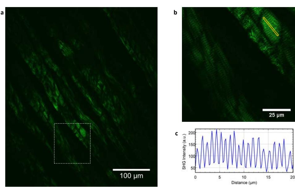
The work shows that widefield SHG microscopy provides sub-micron resolution over a large imaging area. The setup will be used to optimize novel high-repetition-rate amplified laser sources for nonlinear microscopy.