
Two-dimensional (2D) optical spectroscopy is an analogue to multi-dimensional techniques of nuclear magnetic resonance (NMR) as the photon-echo (PE) signal is obtained in a four-wave mixing experiment using ultra-short laser pulses in visible or IR regions. Photon echo signal is then spectrally resolved and plotted against two frequency axis corresponding to two delay times between the laser pulses. 2D PE spectroscopy have been used to probe dynamical phenomena in various multichromophoric complexes. It was the key tool of Engel et al. in proving a wave-like energy transfer within a photosynthetic light-harvesting Fenna – Matthews – Olson (FMO) complex [1].
Theoretical simulations of 2D spectra are computationally expensive and do not scale linearly with a size of the system that is being modeled. The systems that are being investigated most are relatively small. The FMO complex, for example, consists of only 7 chromophores [2]. Here we present simulations of 2D spectra of a much bigger system - J-aggregates consisting of 64 chromophores. Parallel computing on a multi-processor computer cluster had to be implemented.
Molecular aggregates (J-aggregates) are macroscopic clusters of molecules with intermolecular spacing intermediate between crystal lattice and isolated molecules. The scientific interest on J-aggregates is due to the fact that they possess a very narrow absorption band, named the J-band that is red shifted relatively to a monomer band. It is possible that some form of J-aggregates could be used in constructing artificial light harvesting complexes [3].
In this work simulations of 2D spectra of linear J-aggregates of pseudoisocyanine (PIC), 1,1'-diethyl-2,2'-cyanide at 77 K are presented. Many ideas about aggregate structure have been put forward. Here, the spatial configuration model of PIC molecules proposed by Fidder [4] is utilized.
On the left side of Fig. 1 a simulation of 2D spectra at population time $T = 0$ fs of one elementary cell of J-aggregates (4 chromophores in Fidder's model) is presented. As it is expected, the diagonal of 2D spectra mirrors linear absorption spectra. The J-band can be clearly seen at the lower left corner of the spectra at the diagonal. Other features at the diagonal represent to the third and fourth exciton state respectively. As it is expected, they have much smaller amplitudes than the J-band. If the transition dipoles were all parallel, only the J-band would be visible at the diagonal. Cross-peaks that are present at the spectra are due to non zero coupling between the monomers. It should be noted, that ability to see cross-peaks is one of the biggest advantages of 2D spectroscopy compared to linear spectroscopy. The negative peaks are caused by excited state absorption while positive peaks are caused by ground state bleaching and excited state emission.
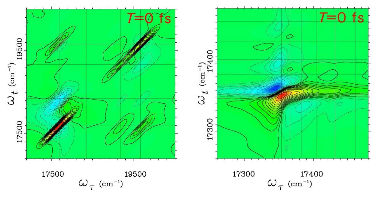
A 2D spectra at population time $T = 0$ fs of J-aggregates consisting of 64 chromophores is shown at th right side of Fig. 1. Because the J-band is getting narrower with increasing aggregate size, only the J-band region of the spectra is presented. The complexity of this spectra can be explained by the fact that visible elements are a superposition of several elements. Still, the positive J-band peak is dominant. The strongest negative peak is much lower here and it changes the form of the J-band peak. Three other negative peaks are visible above the J-band. As population relaxation does not play any role when $T = 0$ fs, these peaks are solely due to excited state absorption.