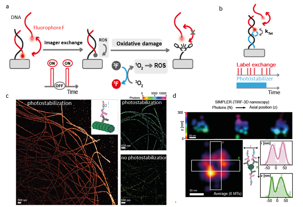
DNA-PAINT is a single-molecule localization microscopy technique, relying on transient hybridization of fluorescently labeled single-stranded DNA imager strands to complementary docking strands on target molecules [1]. During acquisition, docking sites are imaged over the course of multiple binding, dissociation and photobleaching events. Through constant imager strand exchange, the limited photon budget of a single fluorophore is circumvented, making it possible to extract super-resolution images at high laser illumination intensities. Over long periods of continuous high-duty cycle excitation of fluorophores, DNA-PAINT binding sites can, however, be depleted [2]. Fluorophores in triplet excited states may generate singlet oxygen and downstream reactive oxygen species (ROS), damaging the docking sites and labeled target structures (Figure 1a). The use of triplet state quenchers (TSQ) and enzymatic scavenging systems is further limited to systems insensitive to pH change or high additive concentration. Inspired by fluorophore regeneration and self-repair mechanisms, we link the TSQ cyclooctatetraene to a DNA sequence [3], [4]. This photostabilizer strand binds directly next to the imager at the docking site, thereby allowing for self-regeneration and programmed exchange (Figure 1b). The presented contribution shows how this approach can increase the accessible photon budget. The method is characterized in a DNA origami model structure and applied to image microtubules in fixed cells. The improved longevity of DNA-PAINT docking sites is shown and the impact of photostabilizer strand regeneration is explored. The ability to mix and match optimal photostabilizer/dye pairs in this modular approach could be beneficial e.g., for multi-color measurements in structural biology, which often require multiple rounds of imaging, while preserving structural integrity of the sample.(Figure 1c,d)