
Electroporation is a technique to increase cell permeability by applying pulsed electric field (PEF) on cells. Electroporation is typically performed using commercially available cuvettes, nevertheless, it can also be performed at the microscale using a microfluidic chip. In this way, cells are exposed to more uniform electric field, favorable chemical environment, heat dissipates faster [1]. Microfluidic chips are usually fabricated using soft-lithography technique with polydimethylsiloxane (PDMS) as a polymer. Though PDMS is biocompatible and has good optical transparency, its main disadvantages are small molecule adsorption and susceptibility to mechanical stress [2]. Off-stoichiometry thiol–ene (OSTE) can be used as an alternative polymer in microchip fabrication overcoming those disadvantages and retaining the advantages of PDMS [3].
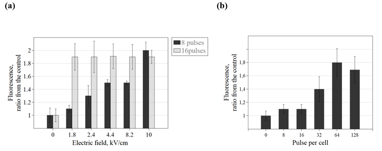
Our laboratory has fabricated the OSTE-based microfluidic chip (Fig. 1). Our goal is to test if the microchip is suitable for mammalian cells electroporation. We performed stop-flow and continuous flow electroporation of rat glioma C6 cell line in the microchip. Cell permeability and viability were evaluated using fluorescent spectrophotometry with DAPI and trypan blue test, respectively. Stop-flow electroporation showed an increase of cell permeability to DAPI with increasing electric field from 1.8 kV/cm to 10 kV/cm with 8 pulses, meanwhile with 16 pulses cell permeability has reached the maximum at 1.8 kV/cm. (Fig. 2 (a)). The viability was not influenced with 8 pulses at all tested electric field strengths. In continuous flow electroporation at 1.8 kV/cm, the same permeability increase was reached with 64 and 128 pulses per cell (Fig. 2 (b)). To conclude, the fabricated microchip is suitable for the PEF treatment of mammalian cells. 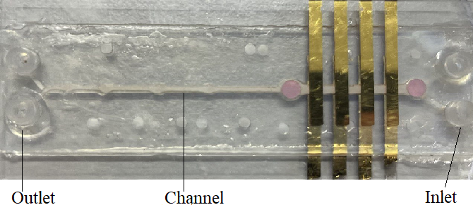
MAMMALIAN CELLS ELECTROPORATION IN THE MICROFLUIDIC CHIP
Agne Damarackaite1, 2, Neringa Bakute1, Arunas Stirke1
1 Center for Physical Science and Technology, Department of Functional Materials and Electronics, Sauletekio av. 3, LT-10257 Vilnius
2 Life Sciences Center, Vilnius University, Sauletekio av. 7, LT-10257 Vilnius
[1] Campelo, S.N. et al. (2023) ‘Recent advancements in electroporation technologies: From bench to Clinic’, Annual Review of Biomedical Engineering, 25(1), pp. 77–100. doi:10.1146/annurev-bioeng-110220-023800.
[2] Wong, I. and Ho, C.-M. (2009) ‘Surface molecular property modifications for Poly(dimethylsiloxane) (PDMS) based microfluidic devices’, Microfluidics and Nanofluidics, 7(3). doi:10.1007/s10404-009-0443-4.
[3] Carlborg, C.F. et al. (2011) ‘Beyond PDMS: Off-stoichiometry thiol–ENE (OSTE) based soft lithography for rapid prototyping of microfluidic devices’, Lab on a Chip, 11(18), p. 3136. doi:10.1039/c1lc20388f.