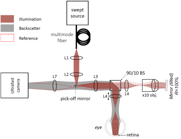
Optical coherence tomography (OCT) is a non-invasive interferometric imaging technique. It can be applied in various situations, such as in-vivo retinal imaging for damage detection, diagnosing and monitoring retinal diseases, imaging deeper layers of human fingerprints, detecting sub-surface defects in optical glasses and etc. However, the spatial resolution, imaging speed and depth is still an issue in OCT. Recently, to speed up OCT imaging, Fourier-domain Full-Field OCT (FD-FF-OCT) has been introduced that uses a swept laser source and a super-fast camera to parallelize signal acquisition. In order to efficiently reduce coherence crosstalk noise in OCT images a multimode fiber can be implemented [1]. Such a system enabled us to visualize such hard-to-image retinal layers with 5µm spatial resolution.
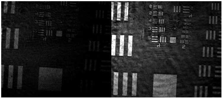
The core of interferometer – beamsplitter is typically 50/50 and wastes almost 75% of light. Light going to the reference arm is practically all lost there because of the strong attenuation necessary when imaging biological tissue. Moreover, half of the light is sent back to the laser source instead of camera. However, by incorporating a small 45° pick-off mirror we were able to use 90/10 beamsplitter (Fig. 1). Such so called high-throughput dark-field (HTDF) [2] FD-FF-OCT configuration [2] allowed us to send more light on the sample and obtain most of the photons with the camera. With this HTDF implementation in our retinal FD-FF-OCT system we have demonstrated 3.5 times increase in signal on a USAF target sample mounted behind a scattering layer.