Zwitterionic meso-tetra(4-sulfonatophenyl) porphyrin and a number of its derivatives self-assemble into tubular aggregates. These aggregates are both found occurring in nature, such as most prominently in the chlorosomes of green bacteria, but can also be synthesized in a laboratory setting. These aggregates have already found use in areas such as photodynamic therapy (PDT) and optoelectronics.[1]
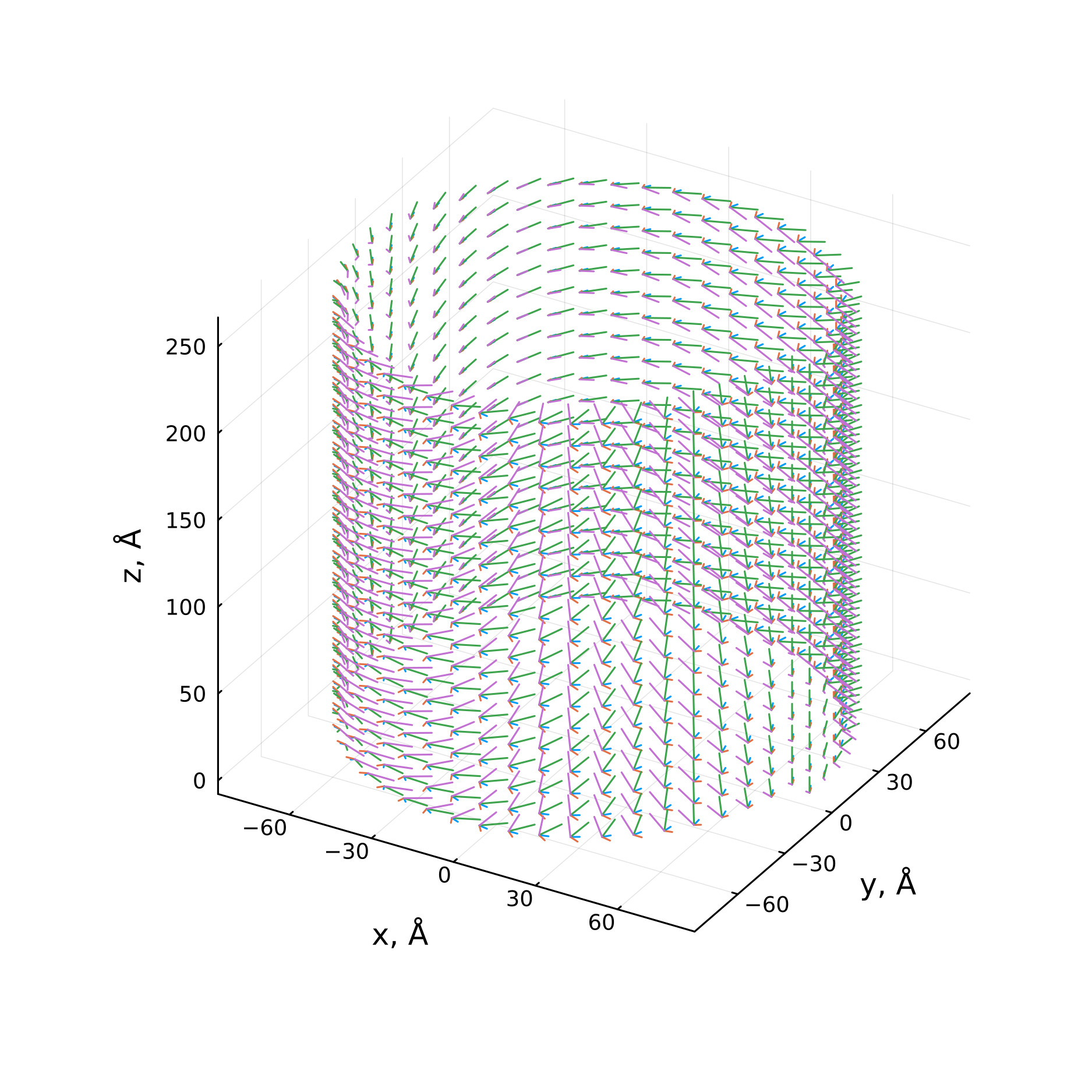
Porphine aggregates have unique optical properties, which can be seen in their absorption and circular dichroism spectra in the range of visible light. It is important to understand the optical responses of these aggregates to better know how to apply them. To achieve this an idealised mathematical model was used, where each molecule in the aggregate was associated with a single point, and arranging them in a helical structure with each point having four optical transition dipole vectors assigned to it. The spectra themselves were calculated by finding the exciton states of the aggregate. These states were found by solving the stationary Schroedinger equation with a hamiltonian constructed with the interactions between each moleculeś respective dipole vectors as its off-diagonal elements. These were then used to calculate both the absorption and circular dichroism spectra of the idealised tube.
Using this relatively simple model we were able to calculate spectra that closely aligns with experiment in its shape and peak positions in both the B and Q bands. To achieve this, many different configurations of dipole vectors and molecules of the aggregate were tested in order to find which closest resembled the experiment. Moreover, taking into account that even a relatively short tube contained thousands of molecules, computational resource use was also taken into account. To that end, the length of the aggregate, adequate to get the highest possible accuracy while also taking up the minimal amount of computation time, was determined.