
Colloidal semiconductor nanocrystals, often referred to as “quantum dots” (QD), have attracted considerable interest in cell labelling due to their unique size-dependent optical properties and high emission stability against bleaching. These unique properties allow them to be used in many applications such as molecular biology, hybrid nanobiosensors and drug delivery systems. Tuneable emission wavelength, broad absorption and sharp emission spectra, high quantum yield (QY), resistance to chemical degradation, versatility in surface modification makes QD very promising fluorescent markers. However, QD synthesized in organic solvents are insoluble in water which is a necessity for biological application and thus further modification is required.
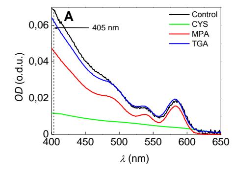
QD solubilisation requires replacing hydrophobic trioctylphosphine oxide (TOPO) surface with a hydrophilic coating. In this work CdSe(ZnS)-TOPO QD surface was modified using thiol group bifunctional ligands: 2-mercaptopropionic acid (MPA), mercaptoacetatic acid (TGA) and cysteamine (CYS). Then the effects on optical properties of QD were observed using spectroscopic methods. Differences in absorption and PL spectra after modification are presented in Fig.1A,B respectively. The decrease of absorbance at the interval ranging from 400 up to 630 nm was detected in both QD-MPA and QD-CYS solutions (Fig.1A).
QD-MPA and QD-TGA photoluminescence (PL) intensity increased in comparison with QD-TOPO (control) while the PL intensity for QD-TGA slightly decreased (Fig.1B). Also a red shift was visible for QD-CYS in PL spectra. QD PL spectral position depends on nanoparticle size, therefore the red shift of QD-CYS might be caused by particle aggregation. The increased QD-CYS PL intensity compared with QD-TOPO implies that the surface coating of QD with CYS molecules the quantum yield of PL but reduces the stability.
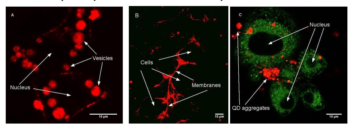
From confocal microscopy experiments was observed, that different ligands on QD surface could determine these nanoparticles uptake and localisation in cells (Fig. 2). QD coated with MPA access the cells and accumulate in vesicles, which after 24 h. of incubation fuse each other into multivesicular bodies. QD-TGA do not pass cells plasma membrane and stuck within also marking membrane structures. QD-CYS aggregates in cell media and do not internalize into cells.
All facts considered, it can be stated that QD properties highly depend on surface chemistry and coating quality. Using surface coating ligands with different functional groups greatly alters both photophysical and biological QD properties. This understanding opens a path for better QD functionalization which allows controlling their localization in cells. In this manner, a new powerful tool for cell probing and biomedicine application could be created.