
Structured monolayers of organic molecules can be a useful tool in controlled cell growth and adhesion experiments. Genome and proteome analysis is another area where such microarrays can be used. Naturally occurring chemical gradients are important for cell growth, adhesion and other biological processes. To study them in a lab, one needs to create controlled chemical gradients [1]. Most of the methods that have been applied to date have disadvantages which prevent efficient manufacturing of complex structures that maintain the function of the biomolecules. D. Rhinow and N.A. Hampp [2] proposed a method of submerged laser ablation which allows fabrication of complex multimolecular self-assembled monolayer (SAM) structures on gold surfaces from various molecules with surface-selective thiol groups. The aim of this work was to investigate the effect of laser radiation on thiol monolayers and to fabricate structures made up of self-assembled monolayers terminated with different functional groups.
In our experiments, a SAM (for instance, HS-C11-EG3-OH) is grown from solution on a gold-covered silicon chips. Initially, the experiments were performed with the specimen put into a small Petri dish filled with ethanolic HS-C11-EG3-biotin solution and covered with glass. Later, a special fluid cell was designed. It makes it possible to change the solutions without moving the specimen, so structures from different molecules (HS-C11-EG3-biotin and HS-C15-COOH) or different concentrations of the same molecule (HS-C11-EG3-biotin) could be formed with greater precision. Pulsed laser NL 640 (Ekspla) was used as irradiation source (wavelength of 532 nm, with an impulse duration of approximately 10 ns). The laser beam was directed using galvanometric scanner, focused using a flat-field lens (100 mm). After sample irradiation, the obtained microstructures were visualized using streptavidin-conjugated quantum dots which selectively bind to biotin groups. The –COOH areas were visualized via non-specific binding of the same quantum dots (an epifluorescence microscope Olympus BX51 was used).
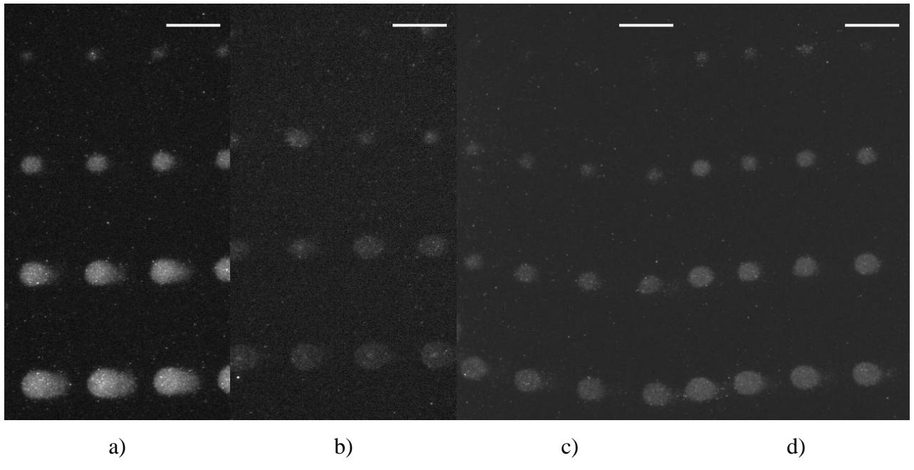
When the HS-C11-EG3-OH SAM is irradiated, the gold-sulfur bonds are broken, molecules (HS-C11-EG3-biotin or HS-C15-COOH) adsorb from the solution and form a new monolayer. Quantum-dot-conjugates revealed that HS-C11-EG3-biotin and HS-C15-COOH molecules form fluorescent spots whose radii depend on the dose of irradiation and laser fluence (Fig. 1 a, b).
Using the fluid cell, the formed structures had a more regular circular shape than those fabricated using a Petri dish.
To check whether, in principle, it is possible to form gradient SAM structures using this method, we performed experiments with HS-C11-EG3-OH SAM in ethanolic HS-C11-EG3-biotin / HS-C11-EG3-OH solutions of different molar ratios. We found that a 70mol% HS-C11-EG3-biotin solution produces more intense fluoresescence of the formed structures than 10mol% HS-C11-EG3-biotin solution, which suggests higher concentration of HS-C11-EG3-biotin (Fig. 1 c, d).
In conclusion, we have confirmed that by irradiating self-assembled thiol monolayer on a gold surface we can form monolayer structures from different molecules. Our experiments show that pulsed laser technologies and SAM chemistry can be integrated to produce biological microarrays. In particular, SAM gradients can be obtained for use in cell growth and adhesion experiments.