
Apoptosis, or programmed cell death, is a physiological cell process essential for normal tissues. It is associated with mitochondrial membrane depolarization and the loss of the electrochemical gradient on the inner mitochondrial membrane. Excessive calcium influx is supposed to be a typical cell death stimulus where mitochondrial membrane depolarization precedes the cytochrome c release. It has been also observed that the inner mitochondrial membrane may undergo a transient hyperpolarization and rapid matrix alkalinization in response to various stimuli during apoptosis. Eventually hyperpolarization and matrix alkalinization are followed by mitochondrial membrane depolarization and equilibration of mitochondrial and cytosolic pH, leading to mitochondrial permeability transition pore opening [1]. Cytoplasmic acidification is required for pro-apoptotic proteins caspases to function properly [2]. Furthermore, the loss of the mitochondrial membrane potential results in the production of reactive oxygen species (ROS) [3]. Most anticancer drugs kill their target cells at least in part through the generation of elevated amounts of intracellular ROS. It is worth mentioning that cytosolic pH values in tumors are above normal levels, which is likely to be due to the activation of one of the major cytosolic pH regulator Na+/H+ exchanger (NHE) in cancer cells. Activation of NHE may lead to inhibition of apoptosis and provide cancer cells survival [2]. Recent findings indicate that either the presence of excessive ROS or the collapse of the mitochondrial membrane potential opens the mitochondrial permeability transition pore, which is accompanied by the release of pro-apoptotic molecules such as cytochrome c into the cytoplasm. Understanding the molecular basis of electron transport chain functioning in tumors will lead to the investigation of novel cancer cell death activation mechanisms by apoptotic signalling pathways controlled by mitochondria and will favour the development of more effective therapies that selectively kill cancer cells with little or no harm to normal cells.
In our investigations the production of reactive oxygen species in human larynx carcinoma cells HEp-2 was observed. In order to reveal possible sources of ROS (HADPH oxidase or mitochondria) the influence of external pH and [Ca2+] on the ROS production in HEp-2 cells was investigated. External calcium and pH did not appear to change the ROS production revealing that HADPH oxidase was unlikely to be a source of the ROS production in HEp-2 cells. The major sites of superoxide formation within the mitochondrial respiratory chain are linked to respiratory complexes I and III [4]. In our experiments both rotenone (complex I inhibitor) and antimycin A (complex III inhibitor) were ascertained to enhance the ROS production in HEp-2 cells. Superoxide production by complex I is supposed to occur during the reverse electron transport (RET) from succinate to NAD+ and during the forward electron transport (FET), the former being faster than the latter. Recent investigations have led to the conclusion that rotenone enhances the ROS formation during the forward electron transport and inhibits it during the reverse electron transport [4]. Moreover, the RET-induced ROS production is regulated by the amplitude of the mitochondrial electrical membrane potential so that a 10% decrease in the membrane potential inhibits the ROS production by 90%. The inhibition of electron transport by antimycin A causes a collapse of the proton gradient across the mitochondrial inner membrane, thereby breaking down the mitochondrial membrane potential [3] but leading to the ROS formation even after rotenone treatment (Fig.1).
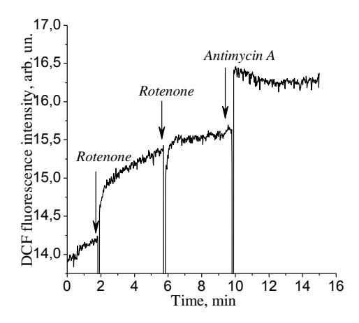
The facts mentioned above indicate that the major source of ROS in HEp-2 cells is mitochondria and the ROS formation is not connected with the reverse electron transport in complex I. Cytosolic pH values in cancer cells are above cytosolic pH values in normal cells. This shift in the cytosolic pH value in cancer cells may lead to a decreased electrochemical gradient on the inner mitochondrial membrane in HEp-2 cells. This, in turn, may result in constant ROS production in HEp-2 cells mitochondria.