Kidney stone disease which causes the formation of urinary stones is very common. However, the formation factors and processes are still not properly understood. Structural and morphological analysis of urinary stones enables to determine the underlying causes of the disease prescribe treatment and prevent recurrences.
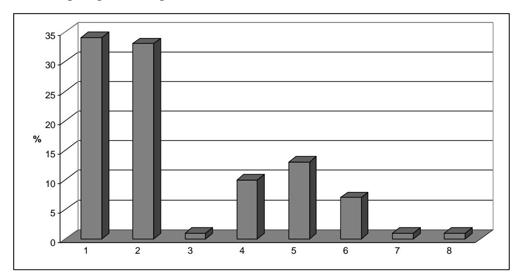
The quantitative and qualitative analysis of urinary calculi is challenging due to their usually small size, fragility and heterogeneous composition. In this work we used two spectroscopical techniques. (I) Fourier transform infrared spectroscopy (FTIR) combined with KBr (potassium bromide) pellet technique allowed us to elucidate chemical composition of the stones. 106 urinary stones received from Vilnius university hospital *Satariskiu klinikos* have been studied in this work. As it can be seen in figure 1, the most abundant components are calcium oxalate and phosphates – apatite and brushite.
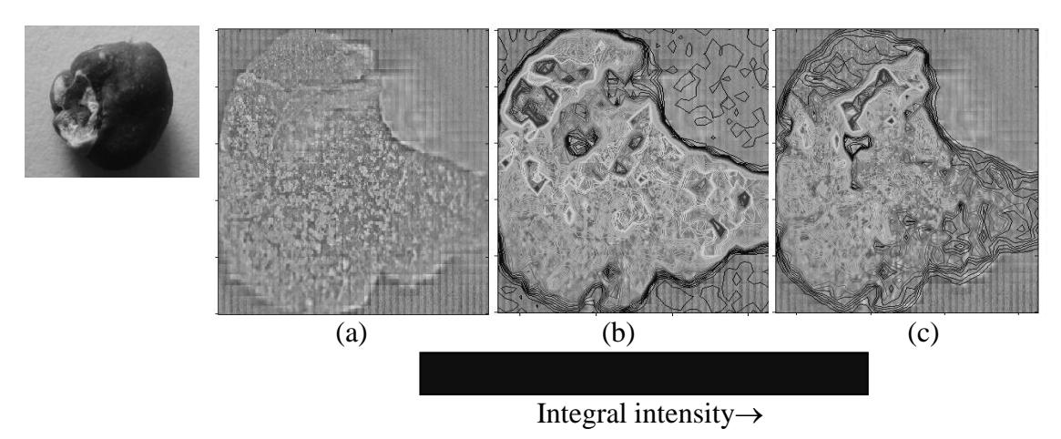
(II) Infrared microspectoscopic surface reflectance imaging allowed us to analyse morphology of the cross-sectioned stones [1]. Integral intensities of the *restrahlen* bands obtained in the specular reflection spectra are directly proportional to the concentration of particular chemical component in the stone (Fig. 2). Distribution of the components in the stones is determined by processes of formation of the stones. In this work, we have experimentally confirmed that stones can be formed in at least two ways: (I) loose stone grows in the urinary system from oversaturated salt solution without fixation to the walls of the system, (II) the stone formation is initiated on the walls. In this case the grow starts with formation of calcium phosphate crystal on the wall of the system. Later, this crystal is being covered by calcium oxalate.