
Semiconductor quantum dots (QDs) are intensively studied for different applications. Size-dependant photoluminescence (PL) spectra, broad and intensive absorption spectra, high photostability and flexible surface modification make QDs attractive as energy donors for different biological fluorophores [1], including porphyrin-type molecules [2]. Porphyrins are typical light activated drugs for the photodynamic therapy of cancer (PDT) with the high singlet oxygen quantum yield and preferential accumulation in tumour. However, the small two-photon (TP) action cross section limits their application in TP-PDT. This problem can be solved by using QDs as energy donors to activate the porphyrin sensitizers.
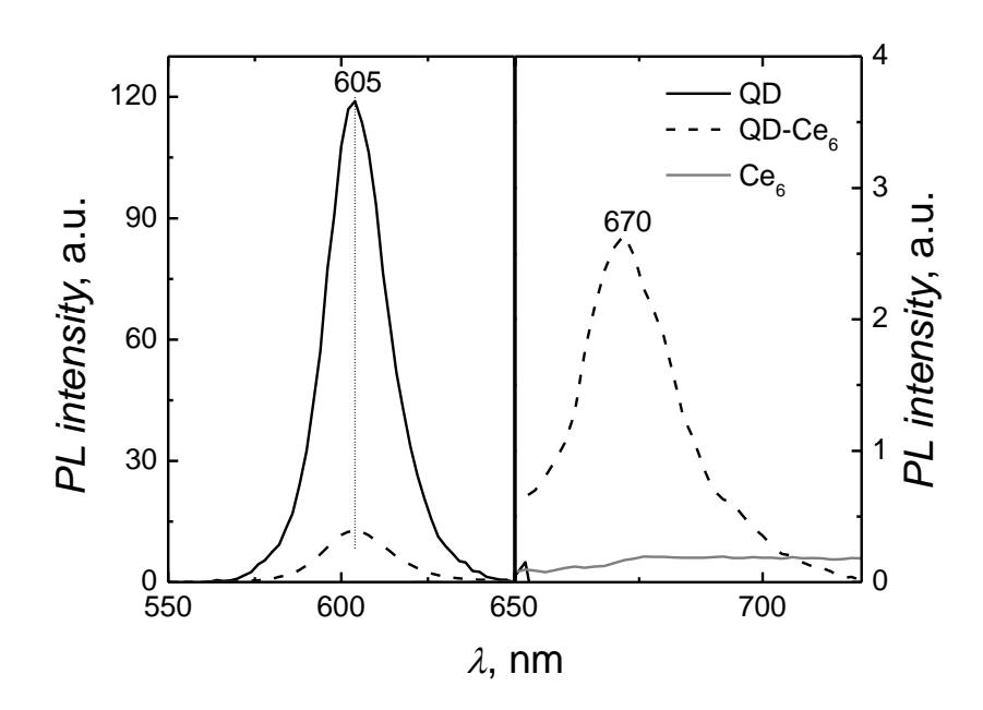
Here we present the study on Forster resonant energy transfer (FRET) from two photon excited CdSe/ZnS QDs to photosensitizer chlorin e6 (Ce6).
For TPE of the samples 810 nm pulsed radiation of Ti-sapphire laser was used. TPE fluorescence spectra of QDs, Ce6 and mixed QD-Ce6 solutions are shown in Fig. 1. The sample of QDs displayed an intensive TPE PL at 605 nm. Meanwhile, the intensity of Ce6 fluorescence at 660 nm, which is usually observed by single-photon excitation, was negligible. The absence of TPE fluorescence indicated the small TP cross section of Ce6. An addition of Ce6 to QDs solution caused the quenching of QDs PL as well the formation of the new fluorescence band at 670 nm was observed. Similar fluorescence band at 670 nm was obtained in QD-Ce6 solutions using single-photon excitation. This band was attributed to Ce6 molecules localized in the lipid-based coating of QDs [3]. Close localization of Ce6 to QD core ensured the effective energy transfer. The hydrophobic environment of QDs coating resulted in 10 nm bathochromic shift of Ce6 fluorescence band. Similar appearance of the intensive fluorescence band at 670 nm in QD-Ce6 solution confirmed the energy transfer from TPE QDs to bound Ce6. The most intensive TPE fluorescence at 670 nm was observed when QD:Ce6 molar ratio was 1:10. The obtained results demonstrated the evidence of indirect PS activation via TPE of QDs, which shows the primary perspectives of using such QD-Ce6 complex for TP-PDT.