
Kidney stone disease affects about 10 percent of the people [1]. Therefore, emerging kidney stones should be found as early as possible. However, usually kidney stones are only found when they have already formed, mainly big and cause severe pain. To suggest a solution to this problem the hypothesis was set: infrared (IR) spectrometry method could be informative enough to investigate human urinary sediment composition and identify the crystals in the urinary sediment which are highly responsible for the kidney stone formation. Urine which is saturated with these materials usually signal about highly anticipated or already existing kidney stone in organism [2]. Continuous monitoring of the patients' urinary sediment composition and its changes could be a way to predict the formation of the kidney stones and then take certain preventive measures to stop kidney stones from growing further.
The urinary sediment samples of Santariskes clinics patients' were tested during the experiment. All spectra were registered using Fourier transform IR spectrometer (FTIR) Bruker Vertex 70. Firstly, centrifuged urine was evaporated until only crystals of urine sediment were left on permeable for IR plate. Then, the IR absorption spectra were registered of these samples. Using the same method the urea sample was prepared and its IR absorption spectrum registered. Recording of the IR absorption spectrum of urea was necessary as it was immediately noticed that urine sediment contains mainly urea. Knowing exactly in which wavelenght values urea molecules absorb, it was possible to reject the influence of urea molecules absorbtion. Then it has been analysed what other low concentration crystal materials could be found in urinary sediment samples.
It was found that excluding urea urinary sediment also contains uric acid. This material is one of the main kidney stones forming substances. Fig. 1. shows uric acid absorbance at 1670 cm-1, 1348 cm-1, 1305 cm-1 wavenumber values. Although uric acid also has characteristic spectral absorption bands in 3160 cm-1 - 2550 cm-1 region, it is likely that because uric acid molecules concentration is low these bands are disguised in the inclined part of the spectrum of the corresponding interval. By forming urine sediment and urea differential spectra, other low concentration crystalline substances were found in the urine sediment.
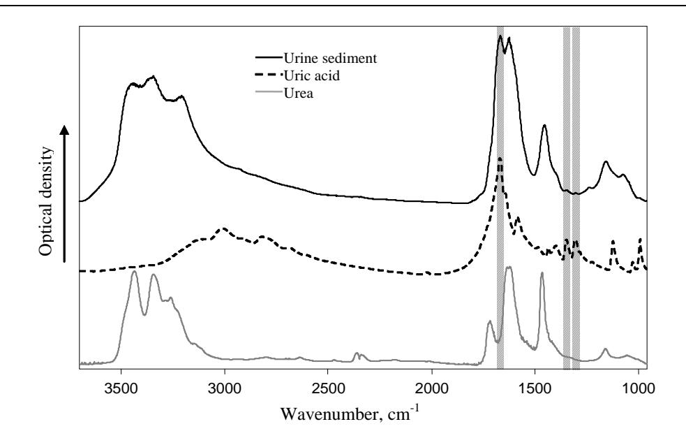
Thus, the selected spectral method is sensitive and informative enough to identify low concentration crystal materials in the urine. Analysis and proper interpretation of patients' urine sediment composition changes could make this method a viable technique for kidney stones surveillance and prevention.