Light microscopy has greatly advanced our understanding of the world on the micro-scale. Various light-matter interactions are used to investigate different physical properties of the specimen, e.g. in bright field microscopy, image contrast is based on absorbance and scattering of light. As developments in optical techniques progress, more specific and contrast mechanisms are used, the higher spatial resolution and higher sensitivity levels are achieved. In particular, the use of non-linear light-matter interactions in microscopy has emerged as a powerful research tool for biology [1]. In the past few years, there had been number of aproaches to combine multiple non-linear contrast mechanisms in one microscope [2,3,4,5].
In this work we present non-linear multimodal optical microscope based on coherent anti-Stokes Raman scattering (CARS) micro-spectrometer [6] and multimodal imaging of some biological objects (i.e. green algae, starch granules). A fundamental beam of picosecond laser operating in a cavity dumping regime (Nd:YVO4, 1064 nm) at the 1MHz repetition rate is used to pump optical parametric generator, which serves as a two-color, signal beam (850-1040 nm) and idler beam (1060-1395 nm), excitation light source. Excitation beams are directed to the standard inverted microscope (Olympus IX71) and focused on the sample with an oil-immersion objective (Olympus, Plan Apochrom., 60×, NA 1.42). This experimental setup can be used for imaging with multiple non-linear contrast mechanisms: coherent anti-Stokes Raman scattering, two-photon excitation fluorescence (TPEF), and second harmonic generation (SHG).
Vibrational origin of CARS signal allows us chemically selective imaging without chemical labeling. In biology, high intensity CARS signals arising from CH2 vibrations are frequently used for lipid membranes, elastin and collagen fibrils imaging. SHG signal arises from non-centrosimetric structures. This characteristic of SHG signal can be used to determine the orientation of molecules in the specimen. TPEF from auto-fluorescent components gives information about the distribution of the fluorophores. Combining information from images of different contrast mechanisms lead to better understanding of the sample under investigation.
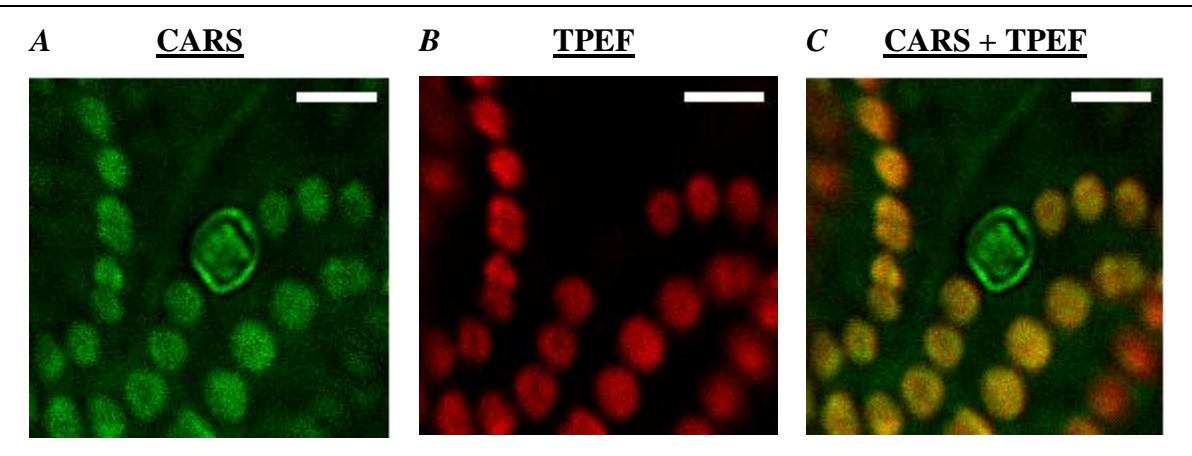
Imaging green algae Nostoc commune* with CARS and TPEF contrast mechanisms are shown in Figure 1. The green algae Nostoc commune* has two types of cells: normal cells which have chlorophyll and heterocyst cells which do not have chlorophyll. Figure 1A shows CARS image tuned to CH2 vibration contrast where both types of cells are visualized. Figure 1B shows TPEF image of chlorophyll fluorescence. In this figure heterocyst cell is not visible.