Cervical cancer is one of the most frequent forms of cancer in women and more than a half of cases of this disease are lethal [1]. Usually methods of cytology and histology are used for diagnostics of the cancer and its pre-cancer states. Unfortunately, these methods can be realized only by qualified specialists in specialized laboratories. So the purpose of our investigation was to search for supplementary or alternative diagnostic methods based on cervical smear material autofluorescence.
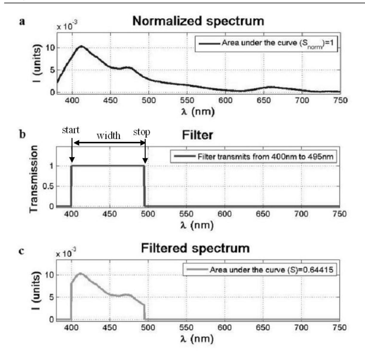
In this research we had samples of cervical smears which were classified into normal and precancerous CIN2+ (cervical intraepithelial neoplasia of 2nd degree or higher) groups according to diagnostic (cytological and histological) tests. Ultraviolet radiation (355nm) of microlaser STA-01-TH was used to excite the samples *in-vitro* and fluorescence spectra were registered. Method of spectra filtration was developed to analyze individual regions of fluorescence spectra. The main procedures of this method are spectra normalisation, rectangular function (i.e. rectangular filter) creation and filtration (Fig.1). This sequence of procedures was repeated 4524 times with different parameters (stat position and width) of filter. Then areas under the curves of filtered spectra were calculated and compared between normal and CIN2+ groups using analysis of ROC curves. Source codes have been developed in order to perform this analysis automatically. In several spectral regions statistical parameter AUC (area under the ROC curve) of 0.71 has been achieved. According to literature [2], this indicates diagnostic value of the test as 'good'.