
Kidney disease is a growing problem. In some countries up to 12 percent of total population suffer from kidney stones and it is still unknown how and why these stones are being formed. It is very important that the causes of stone formation must be understood in order to enable the doctors stopping the disease and preventing its recurrence. Therefore the studies of chemical composition and domain structure of the stones are of the great importance.
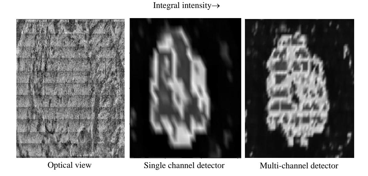
Fourier transform infrared (FTIR) microscopy [1, 2] is an effective method for qualitative and quantitative analysis of the spatial distribution of chemical compounds in kidney stones. In this study Bruker “Hyperion 3000” infrared microscope was used, connected to FTIR spectrometer „Vertex 70“. The data was registered and processed using „Opus 6.5“ software package. The spectra were collected using specular reflectance method. In the analysis of the sample single channel semiconductor mercury-cadmium-telluride (MCT) detector and multi-channel focal plane MCT 64x64 array (FPA) detector were used. Multi-channel detector enables to achieve higher spatial resolution (up to 20 μm) compared to a single channel detector (100 μm). The results showed that chemical maps, created using FPA detector, were more detailed and smaller domain structures were visible (Fig. 1).
These studies also allow concluding that the stones can be formed in at least two different ways. Formation of the stone can be induced by small deposit of one component on the epithelium of urinary tract and a layer of another component forms on it through time. In this particular case stone had calcium oxalate core with uric acid layer formed around it. Stone can also be formed in the oversaturated urine from various mineral salts.