
Application of nanotechnologies in medicine has been one of the most active research fields during the past decade. Various nanoparticles were investigated as promising contrast agents for MRI and X-ray spectroscopy, targeted drug delivery or as photothermal or photodynamic therapy agents. Photoluminescent gold nanoclusters are one of the latest and very promising findings in nanomedicine field because of their remarkable optical properties for a wide range of applications such as sensing, biolabeling and imaging [1]. However, only few studies have been reported on the growth of gold nanoclusters capped with bovine serum albumin (BSA). In this work spatial structure of gold nanoclusters capped with BSA (Au BSA) was investigated using atomic force microscopy (AFM) and quasielastic light scattering techniques.
Au-BSA nanoclusters were synthesized following synthesis conditions published by Xie, et al. [1]. Several different Au to BSA molar ratios were chosen in order to find synthesis conditions for Au-BSA nanoclusters showing the best photoluminescence properties. Further experiments for estimation of spatial structure were performed on Au-BSA nanoclusters synthesized under optimal Au to BSA ratio.
Hydrodynamic diameter of Au-BSA nanoclusters was measured with "ZetaPlus” - particle size measurement equipment based on quasielastic light scattering using approximation of nanoparticles as spheres. Results showed that in aqueous solution Au-BSA nanoclusters were around 9 nm in diameter (Fig.1, b). This is approximately the diameter of the BSA molecule reported in literature [2].
Atomic force microscopy measurements were performed in tapping mode. Samples were prepared on freshly cleaved mica's surface using spin coating technique. Round ~1.3 nm in height and ~30 nm in width structures were observed on the surface (Fig.1, a).
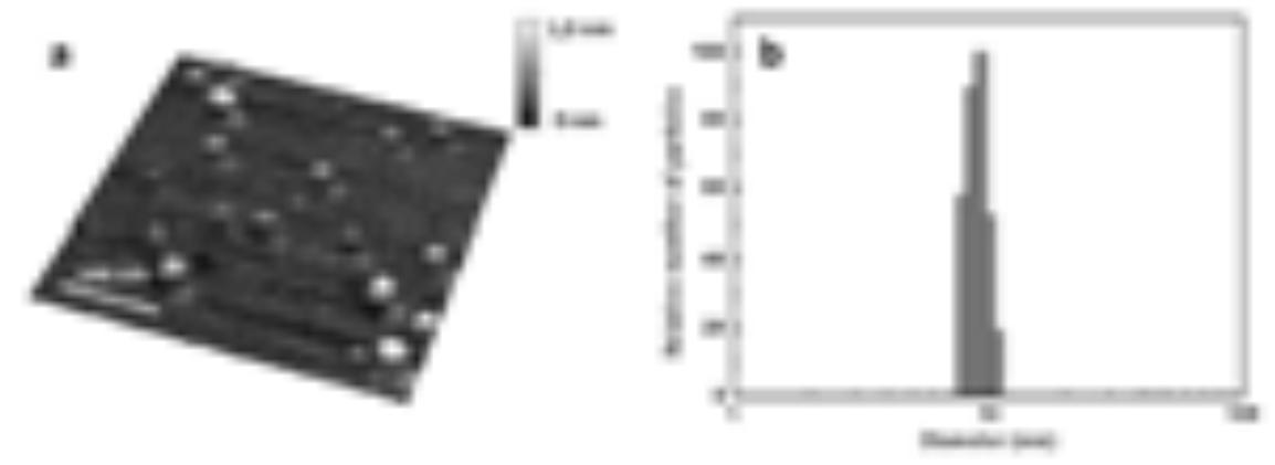
The estimated spatial structure differences could be due to the different environment of Au-BSA nanocluster when different measurement techniques were used. In order to explain these differences the modeling calculations were performed. The spherical shape of the Au-BSA nanoclusters could be suggested in aqueous solution in case of quasielastic light scattering method used. On the surface of mica the spherical structure should be flattened. The change of the spherical shape structure was simulated theoretically by the flattening it till the height of the round structures measured with AFM. The diameter of the flattened ellipsoid was two times smaller than the diameter of round structure estimated from AFM measurements. It seems that change of Au-BSA nanoclusters shape is not only due to environment phase changes but also because of the interaction of BSA with hydrophilic mica's surface which is reported to have strong electrostatic interaction with BSA molecules.
However despite the drastic changes of spatial structure of BSA, gold atoms inside remained bound together and clusters on mica's surface showed typical absorbance and photoluminescence spectra.