
Quantum dots (QD) are fluorescent semiconductor nanoparticles with unique physicochemical properties which make them attractive for early cancer cells diagnostics and therapy. In contrast with organic dyes QD are stable, bright fluorophores with high photoluminescence (PL) quantum yields, narrow PL bands, high absorbance and high resistance to photobleaching and degradation in vivo. PL wavelength of these fluorophores is dependent on their size which is adjustable. However use of QD for cancer cell visualization is complicated because they are not selective probes by themselves.
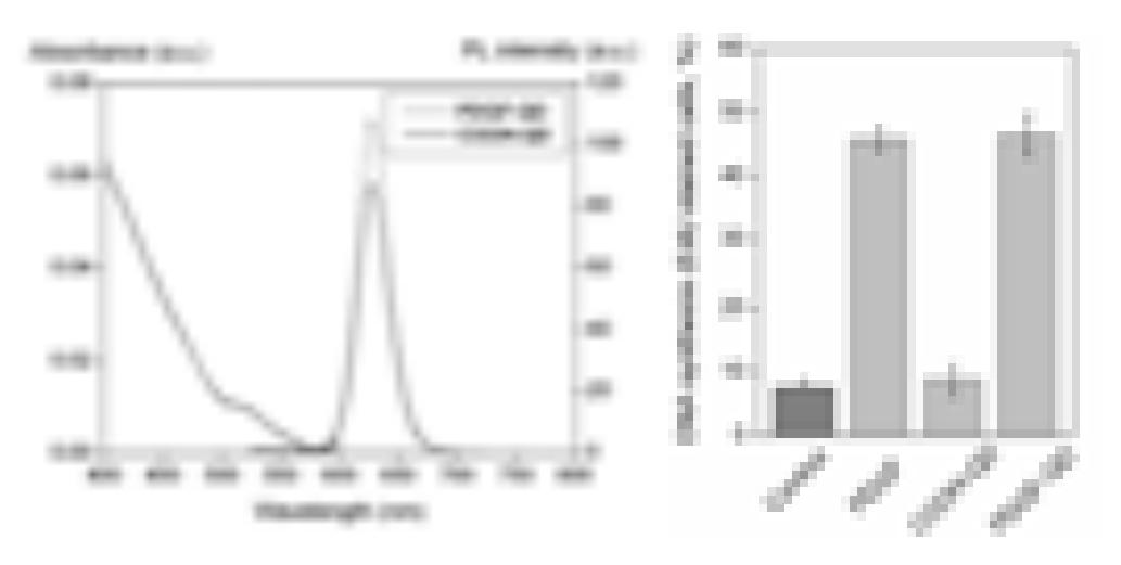
The goal of our study was to prepare selective QD conjugates that could recognize cancer cells. For that purpose platelet derived growth factor (PDGF-BB) was selected. This protein is homodimeric growth factor which binds to platelet derived growth factor receptor (PDGFR) and initiates its dimerization, autophosporylation and activation. This leads to cascade of signaling events in the cells that regulate cell growth, proliferation, differentiation, migration and other processes. To date, PDGF expression has been demonstrated in a number of different solid tumors, from glioblastomas to prostate carcinomas [1]. Characterization of photophysical properties of PDGF targeted QD conjugates (PDGF-QD) was performed by using steady-state spectroscopy. Absorbance and photoluminescence (PL) spectra of none coupled QD and PDGF-QD is presented in Fig. 1A. Both non-coupled and PDGF-QD absorb light in wide spectral region and have PL band peak at 628nm. This shows that surface modification of QD does not change their colloidal stability; however, coupling of PDGF-BB to QD surface slightly increases PL intensity. To evaluate how surface modification of QD with PDGF-BB affects biological properties we investigated their effect on DNA synthesis induction in embryonic mouse fibroblast NIH3T3 cells. Cells were treated with none targeted QD, PDGF-QD and PDGF alone and their proliferation was estimated by confocal microscopy using Click-iT EdU Imaging Kit. Treatment of cells with PDGF or PDGF-QD, but not with none coupled QD, induced DNA synthesis to the similar level (Fig. 1B). Fig 1B shows, that PDGF-QD are capable to elicit potent mitogenic response.
For cancer cells detection it would be necessary that ligand do not cause proliferation of cells. For that purpose PDGF protein was monomerized and coupled to QD. Its validity as biosensor was evaluated by imunoprecipitation and fluorescence methods.