
Nanomedicine is currently an active field. This is because new properties emerge when the size of a matter is reduced from bulk to the nanometre scale. These new properties, including optical, magnetic, electronic, and structural, make nanosized particles (generally 1-100 nm in diameter) very promising for a wide range of biomedical applications such as cellular imaging, molecular diagnostics and targeted therapy depending on the structure, composite and shape of the nanomaterials.

Gold nanoparticles have been brought to the forefront of cancer research in recent years because of their easy synthesis and surface modification, strongly enhanced and tunable optical properties (Fig. 1), as well as excellent biocompatibility feasible for clinic settings. Gold nanoclusters are assemblies of small number (2-30) of gold atoms, typically having the size no bigger than 1 nm. These nanoclusters demonstrate molecule-like properties distinct from bulk metals or atoms. Stabilization of these nanoclusters in aqueous solutions protects them from aggregation. Reduced gold atoms aggregate within the bovine serum albumin (BSA) to form small and large nanoparticles. This protein-directed synthesis of highly fluorescent gold nanoclusters was firstly investigated by [1]. Here based on [1] we investigate optical properties of bovine serum albumin coated gold nanoclusters.
At the moment the process of BSA-gold nanoclusters interactions, photoluminescence and absorbance spectra, temperatures kinetics interpretation or two photon excitation cross sections is very poorly or absolutely unknown. In this report results on spectra, fluorescence kinetics interpretation, two photon cross sections of BSA coated gold nanoclusters will be presented. At the moment there are only few articles investigating the tumor NIR fluorescence imaging in vivo with the ultrasmall gold nanoclusters which exhibits brightly NIR fluorescence, high photostability and contains no toxic elements [1]. The uptake by the RES system in vivo is also much less than other nanomaterial due to the ultrasmall hydrodinamic size.
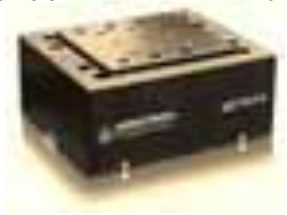
Looking forward gold nanoparticles are shown to be good to carry out non invasive cell optoporation (nanofothothermolysis). Using femtosecond laser system PHAROS (Light Conversion Ltd.) operating at $\lambda=1030$ nm and delivering pulses of 270 fs at 200 kHz repetition rate and sample mounted on a three-axial motion platform (Fig. 2), controlled with the SCA software (Altechna Ltd.) precise cell optoporation will be caried out. So scientists believe that the biocompatible, ultrasmall, NIR gold nanoclusters are promising fluorescent candidates for optical imaging in vivo and related biomedical applications.