
Degeneration is a process occurring due to mechanical loading such as walking, running, bicycling or any other form of intense movement. Treatment of degenerative diseases and injuries is quite challenging. New methods are being developed constantly, for example, gelatin scaffolds are now applied for tissue engineering purposes, more accurately to foster in vivo regeneration of cartilage tissue. The basic aim of the research was to characterize cell-gelatin scaffold interactions using optical tweezing method.
Basic materials that were used for the research: cells and gelatin scaffolds. Experiments were held using mouse fibroblast cell line L-929. They are widely used in many experiment aspects as the fibroblasts are the most common cells of connective tissue in animals and they play an important role in wound healing. Gelatin scaffolds were made after crosslinking procedure of B type gelatins. Type B gelatins are derived from collagen of beef hides and are produced by partial hydrolysis and denaturation of collagen, during which the regular triple helix structure is broken down to form random gelatin coils [1].
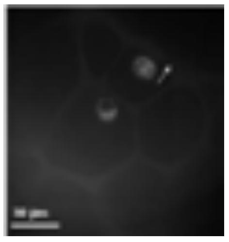
Research has showed, that proliferation of cells in the gelatin scaffolds is very good - cells are growing profusely. Cells were labeled with amphiphilic fluorescent dye with the high value of membrane/water partitioning coefficient. The efficiency of trapping is in general dependent on the differences in refractive indexes of a dielectric object and surrounding media, the laser beam itself (beam characteristics, wavelengths) and the objective properties (numerical aperture, immersion objectives, etc.)[2]. Very promising is that, despite a little higher refractive index of cells ($n=1.35-1.37$) [3], [4] compared to refractive index of surrounding water ($n=1.33$), cells still can be trapped and slightly manipulated, considering of moving, stretching, etc. as it is showed in the picture (Fig. 1).
Force acting on the cell trapped in optical field when moving the cell away from the surface is the force of binding proteins, which bind extracellular matrix and scaffold matrix. If the force of binding proteins exceeds estimated trapping force, then the cell disappears from the trap (Fig. 2).
The total force on an object due to refraction of light is:
$$ \mathbf{F} = \left(\frac{\mathbf{n}}{c}\right) \iint \left(\mathbf{S}_{in} - \mathbf{S}_{out}\right) d\mathbf{A} \tag{1} $$where n – refractive index of surrounding media, c - speed of light, Sin- Poynting vector, which describes the momentum flux of photons entering the object, respectively Sout - momentum flux of photons leaving the object, dA – an element of area normal to S [5]. The integral represents the total power of the light. In real case the maximum efficiency is hardly achieved so the force equation is multiplied with the quality factor Q which depends on the direction in which force is measured. The highest factor is in perpendicular direction according to propagation of laser beam. Eq. 1 becomes then:
$$ \mathbf{F} = \left(\frac{\mathbf{n}}{\mathbf{c}}\right) \cdot \mathbf{W} \cdot \mathbf{Q} \tag{2} $$