
The most convienient way to investigate radiation effects is to use protons or ions beams produced by accelerators. It is because of simplicity to control dose and high probability of double strand breaks (DSBs) in the DNA because of direct ionizing. Free electrons are the key particles to cause lesions of the DNA [1]. But the enegy of these electrons is low and there is no experimental data for that spectrum. Though it exist GEANT 4 – DNA extension where it is possible to simulate protons of energy from 100 eV to 100 MeV and electrons of energy from 8 eV to 1 MeV [2].
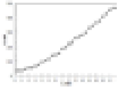
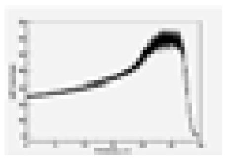
It was calculated that in experimental conditions (100 μm diameter slit [3] for protons exit and 1.6 MeV initial energy) beam radius at intensity FWHM expands to 410 μm(Fig. 1). It was also investigated that enegy of protons at the surface of cells should be 1.075 MeV which means that range of a proton in water is ~30 μm and it is three times more than a cell. To determine dose it was important to know how many energy one proton leaves in a cell. So LET curve was calculated(Fig. 2). Calculations were done with GEANT4 [4, 5] and MCNPX [6].
The main thing doing radiobiological experiments is to deterimine absorbed dose. Faraday cup was used in this particular investigation. Evaluated dose was 200±40 mGy which is near declared hormesis threshold. That is possibly why survival of irradiated cells were 7% better than non – irradiated cells (82±1% VS 75±1%). Cell line MIA PaCa-2 was used in this investigation [7]. Viability was checked with cell counter ADAM.