
Rheumatoid arthritis (RA) is a chronic, inflammatory disease that primarily attacks flexible (synovial) joints. About 1% of the world's population is afflicted by the disease, with women being three times more likely to have it than men. The most common age of onset is between 40 and 50, however it can develop for people of any age. The disease is usually followed by pain and finally it can lead to a loss of mobility and everyday functioning without proper treatment [1]. About 60% of synovial fluid samples collected from patients with arthritis contain calcium pyrophosphate dihydrate (CPPD) or basic calcium phosphate (BCP) crystals [2]. The presence of CPPD or BCP crystals can cause severe joint damage and rapid progression of RA. The relationship between CPPD and BCP and RA is still an unsolved puzzle [3]. Infrared (IR) microspectroscopy method for the investigation of synovial fluid has the potential to provide early diagnosis of the RA and also to grade the severity of the illness in arthritis patients by analyzing the chemical structure of crystals found in the affected joints[2, 4].
The sample of synovial fluid was collected from a patient suffering from active rheumatoid arthritis. 50 μl of synovial fluid was left to dry on a CaF2 optical window. Then the spectra from different regions of dried synovial fluid film were collected: for areas dense with small crystals and for areas where no crystals were deposited. The measurements have been done over the 890-4000 cm-1 spectral region using a Bruker Vertex 70 spectrometer with an integrated IR microscope Hyperion 3000.
The summarized findings can be seen in Fig. 1. Most obvious IR absorption spectral band at 1044 cm-1 appears in the spectra of synovial fluid area which is dense with small crystals. Those are most likely to be CPPD crystals having absorption bands in the region shown in Fig. 1(b). The same absorption bands do not appear in the spectra of areas which are not affected by crystals accumulation.
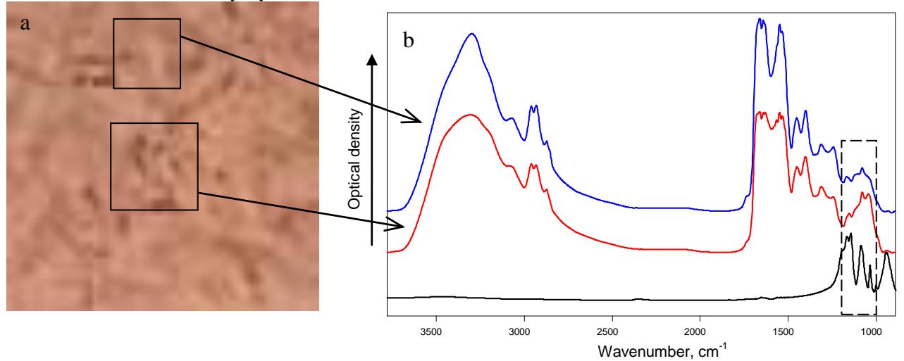
Our results show that infrared spectroscopy is able to supply information about chemical constitution of crystals of synovial fluid. This method is superior to optical imaging – conventional method used in contemporary medicine. Information about chemical structure of the crystals is essential to help the patient's physicians ascribe the appropriate treatment. Infrared spectral microscopy could be a possible technique to diagnose arthritis – it can be applied to investigate the crystals responsible for degeneration of joint tissue.