
The classical scheme of tissue oxygenation is based on the fact that oxygen delivery is taken in through by the capillary network [1]. In accordance with modern experimental measurements of the longitudinal and radial oxygen gradients in blood vessels, it was shown that oxygenation in tissues is being provided, first and foremost, with arterioles and secondarily with capillary. The velocity of transmural oxygenation for these tissues is so high, that couldn't be explained by the diffusive concept, because oxygen permeability of the arteriole wall in vivo was much more higher than in vitro. In accordance with convective mechanism of tissue oxygenation concept the vectorial water and oxygen mass transfer between vessels and water compartments in tissues is taken in through by aquaporins [2].

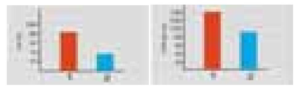
We have installed a sensor on the rabbit's carotid in vivo and we measured pO2 level on it (before and after ligaturing) (fig 1). We have estimated oxygen permeability of the part of rabbit's ligatured carotid in internal and external directions. The carotid was cut, unfolded and placed on the muscle, at first, with an internal surface on the top, secondary, with an external one. We had measured oxygen mass transfer trough the vessel's wall to the muscle, oxygen consumption velocities of the muscle and of internal and external surfaces of the carotid's wall. The carotid's wall permeability is a difference between oxygen transfer and carotid's wall cells oxygen consumption.
On the external surface of artery it was detected physiologically high levels of pO2 (81 mm Hg). Ligaturing leads to the pressure decreasing (fig.2). It is obvious that transmural oxygen mass transfer is taking place. It determines high levels of pO2 on external surface of carotid's wall and could take part in nearest tissues oxygenation. It was developed that oxygen mass transferring from inside to outside the vessel is higher than backwards (fig. 3).