
Quantum dots (QDs) - semiconductor nanometer-size particles are promising fluorescent markers. Compared with organic dyes, QDs have a narrow and tunable emission band, broad absorption and sharp emission spectra, high brightness, enhanced photostability, resistance to chemical degradation and photobleaching [1]. The research on QDs is important in order to provide safe and effective biosensing materials for medicine [2].
In this study, absorption and steady state spectroscopy measurements were performed on CdSe/ZnS quantum dots coated with thioglycolic acid (TGA) in biological model system. Stability, photostability and spectral properties of quantum dots were investigated in aqueous media to reveal the effects of QDs interaction with bovine serum albumin (BSA) (Albumin, V fraction, M=69000 g/mol, Carl Roth GmbH, Germany).
It was found that protein increases photoluminescence (PL) intensity of QDs straight after its addition (Fig. 1a). Interaction of quantum dots with BSA caused a blue shift of photoluminescence band from 548 nm to 545 nm. PL intensity of CdSe/Zn-TGA quantum dots solution with protein was the most intensive on the second day. However, the PL intensity of QDs without BSA was decreased (Fig. 1b). It seems that protein interaction with quantum dots increases a photoluminescence quantum yield and makes them more stable. This effect could be resulted by formation of a new layer of quantum dot coating [3].
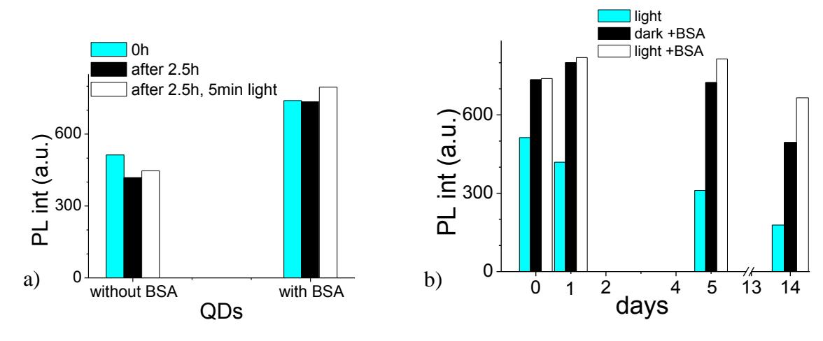
Samples in quartz cuvette were exposed to laser light (532 nm, 35 mW/cm2) to measure photostability. A uniform decrease in PL intensity occurred during 2.5 h in QDs solution, but for QDs with BSA this decrease was not observed (Fig. 1a). Laser light immediately increased PL intensity of QDs and also made samples more stable. As figure 1b shows, the PL intensity of the samples, which were exposed to light, decreases less when all samples are kept in the dark. Also, photoluminescence intensity of QDs decreased less in exposed solution with BSA during 14 days. The CdSe/Zn-TGA quantum dots in aqueous media were the most stable with bovine serum albumin and exposed to laser light. It seems that both light expose and protein can be used to enhance QDs the photoluminescence intensity.