
Kidney stone disease is a common disease with prevalence for about 10% of human population [1]. Most kidney stones formers have disorders either in metabolism and excretion of stone forming materials or in promoters and inhibitors of their crystallization [2, 3]. As crystalline components coalesce in clusters they start to form solid kidney stones. The early identification of kidney stone forming crystals in human urine is essential for taking early preventive actions for kidney stone disease [4]. Usually optical microscopy is employed for the identification of the chemical composition of such crystals or crystal clusters, and such identification is based on their morphological structure. However, the examination of optical view is based on laboratory personnel skills and has significant probability of misinterpreting the chemical composition of urinary crystals. Infrared spectroscopic microscopy (mFTIR) could be used as a more reliable method to correctly identify chemical composition of urinary crystalline components.
Urine samples were taken just before kidney stone removal surgery, centrifuged and filtered on ashless filters for 24 hours to separate urea and water from bigger urinary crystals. The latter were transferred on IR transparent CaF2 (3800 – 890 cm-1 spectral region) or ZnSe (3800 – 600 cm-1 spectral region) window and examined by Bruker Hyperion 3000 IR microscope.
We examined 72 kidney stones patients' of Vilnius Santariskes clinics and 5 healthy persons' urinary sediment. In order to evaluate the correlation between urinary crystals and kidney stone chemical composition of the same patient, both were investigated. In 52% cases, patients with kidney stone disease had the same chemical composition of crystals in their urine as their kidney stone chemical composition. It was found no kidney stones forming sediment in the urine of healthy people.
There were found both regular shape crystals and some undefined shape crystals or crystal clusters. Optical images of some typical examples of urinary crystals and their infrared spectra are presented in Fig. 1. Whatever morphological shapes of the crystals, we were able to discover main kidney stones forming materials: calcium oxalate, uric acid, hydroxyapatite, brushite, struvite – as monocrystals or in crystal clusters. Drug metabolites and proteins were also found as sediments.
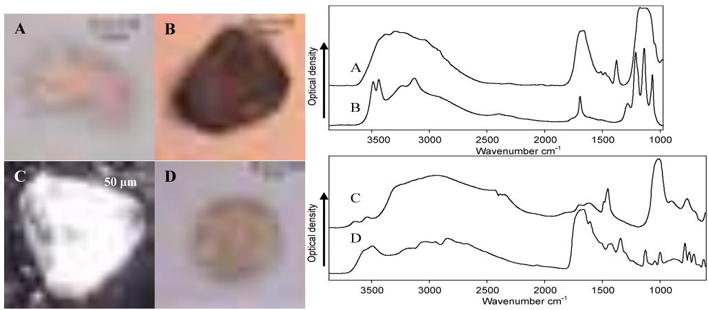
Our experiments revealed that mFTIR method is informative enough, reliable and easy to implement in order to define the chemical composition of urinary crystals no matter their morphological structure. Also, the correlation between chemical composition of both, kidney stones and urinary sediments of the same patients suggests, that examination of urinary sediments by mFTIR spectroscopy can be considered as a relevant technique for early identification of kidney stones and determination of appropriate actions to prevent their formation.