
Formation of kidney stones is very painful urinary pathology. Approximately 8-15% of people in Europe suffer from urinary stones disease. 65 different organic and inorganic chemical compounds can be found in urinary stones [1]. Oxalatic and uratic stones are most common types of kidney stones. Every piece of information about composition and formation of kidney stones is important in searching ways for treatment. Raman spectroscopy is very informative method for structural studies of molecular substances. Unfortunately, application of this method is very limited in case of "real" non-purified substances due to strong fluorescence background appearing in Raman spectra of such compounds. In this work an attempt was made to apply Raman spectroscopy for uratic kidney stones, which are problematic for Raman spectroscopy samples due to strong fluorescence background.
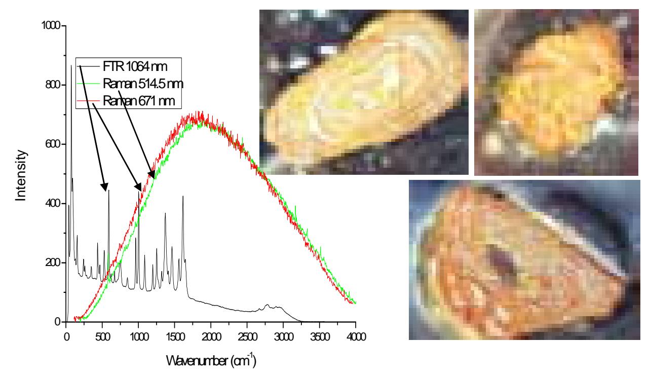
10 uratic stones, removed surgically or by ultrasound technique from urological patients of “Santariškių klinikos” were taken for structural studies by means of Raman spectroscopy. The samples for Raman experiments were prepared in 3 ways: (I) untreated stones (II) cross sectioned stones and (III) powdered stones. Three different lasers (514.5, 671 and 1064 nm) were used for excitation of Raman spectra of the stones, but only in third case (FT-Raman with NIR excitation) the Raman spectrum was not masked by fluorescence background (see fig. 1). FT-Raman spectra were obtained for all 10 uratic stones, which differ in shape, colour, porosity, etc. (see fig. 2). It was found that 8 of them consist from pure uric acid while 2 – from calcium oxalate and uric acid. After close up examination of the stones, which included calcium oxalate it was found that white stripes between calcium oxalate and uratic part of stone is pure calcium oxalate.
Notable, that main method used for determination of kidney stone type in hospitals is optical examination of shape and colour of the stone. These physical properties not always represent chemical composition. To sum up FT-Raman spectroscopy gives more information about structure of kidney stones than traditional optical methods and can be used in medical practice.