A few decades ago it was noticed that cancerous tissue is not homogeneous but has cellular hierarchy and different levels of differentiation [1]. A small subpopulation of cancer cells is associated with tumor regeneration, metastasis, multi-drug resistance. These cells are called cancer stem cells (CSCs) as their properties are similar to normal stem cells [2]. CSCs can initiate tumor growth and give rise to other less aggressive cancer cells. It is very important to define the characteristics of CSCs and to find a new method of treatment targeting exclusively cancer stem cells.
Quantum dots (QDs) are nano-sized crystals made of semiconductor materials. They are advantageous for improved cellular imaging and delivery of active molecules directly to tumors. QDs have broad excitation and narrow fluorescence emission spectra and are more photostable than organic dyes. They can also serve as a platform for biologically active compounds such as antibodies and/or drugs [3].
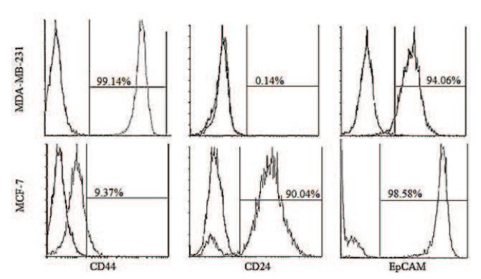
In our study we used two different breast cancer cell lines – MCF-7 and MDA-MB-231. To determine which cell line has more cells potentially to be cancer stem-like ones we performed three different tests. As CSCs are distinguished by better colony formation, resistance to chemotherapy and expression of specific surface molecules, we used plating efficiency, sensitivity to doxorubicin and immunophenotyping with antibodies against CD44, CD24 and EpCAM antigens (Fig.1.). For accumulation studies we used eFluor® 605NC QDs and the conjugate of anti-CD44 and eFluor® 605NC QDs. The photoluminescence of QDs inside cells was registered by Nikon C1si laser scanning confocal microscope.
The results show that the cell line with the subpopulation of CSCs accumulates much more QDs even without the anti-CD44. Although such a connection between accumulation of QDs and CSCs yet remains to be investigated, it might open the way to the elimination of this critical population of cancer cells.