DPN is a scanning-probe-based lithography, in which tip is used to make nano-scale chemical patterns by directly transferring molecules to the surface. It was first introduced in 1999 [1]. This method offers a number of advantages over other nanofabrication methods, such as no need of expensive photolithographic masks, but it also has some issues such as slow patterning speed and accurate control of the material amount on the tip, which dramatically affects the repeatability of the structures.
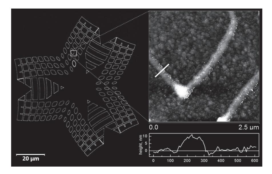
The goal of this study was to explore the possibilities of dip-pen nanolithography (DPN) to fabricate lipid patterns on solid supports. An ink-jet based tip inking [2] was used as a novel method for such a control. Investigation of fluorescent lipid 1, 2-dioleoyl-sn-glycero-3-phosphocholine (DOPC) mixed with 1% 1, 2-dioleoyl-sn-glycero-3-phosphoethanolamine-N-carboxyfluorescein (DOPC-FL) traces on different tips using epifluorescence microscopy showed a reliable repeatability of the material deposited on the tip. After that patterning using JPK scanning probe microscope was done on the silicon dioxide surface in order to investigate how this inking strategy affects DPN patterns. Fabricated patterns investigated by using fluorescence and scanning probe microscopy showed a reasonable repeatability and line widths down to 200 nm on a silicon dioxide surface. Lipids allowed reaching high patterning speeds (up to 100 μm/s). Furthermore, DPN was done on different surfaces such as self-assembled monolayers (SAM's) in order to investigate lipids spreading and behavior on different hydrophobicity surfaces.
Results of this research show that ink-jet material deposition on the tip allows reaching significant improvement over conventional ink well inking and provide possibilities to develop new applications of DPN technology.