Polymer coatings have a great importance in a variety of fields. They are used for drug delivery and biocompatibility of biomedical materials. Thus, it is a must to understand origin of chemical bonds formed between components of multi-polymer layers. Vibrational spectroscopy techniques such as Raman scattering or infrared absorption are known as fast and reliable methods for determination of type of chemical bonding. However, these conventional spectroscopic methods are not sensitive enough for investigation of thin surface films and different approaches must be taken. One possible approach is surface enhanced infrared absorption spectroscopy (SEIRA) and surface enhanced Raman scattering spectroscopy (SERS).
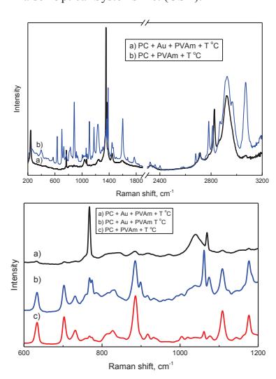
In this work, colloidal solutions of different sizes and shapes of silver and gold nanoparticles were introduced into polymer (polycarbonate (PC), polyethylenimine (PEI), polyvinylamine (PVAm)) interfaces in order to enhance the spectral signals of their reaction products. Three types of polymer systems: PC-Ag(Au)-PC, PC-Ag(Au)-PEI and PC-Ag(Au)-PVAm were analysed. The reaction between polymers was initiated by heating of the films in a vacuum oven or in a variable temperature cell.
The samples for the infrared absorption measurements were prepared by spin coating the polymer solutions on a calcium fluoride (CaF2) substrate. For Raman scattering measurements a PC foil of 250 μm thickness was prepared by pressure heating. The second polymer was brushed on and dried. Colloidal particles were introduced to the system by drying the colloidal solution on the polymer or spin coating the second polymer diluted in the colloidal solution.
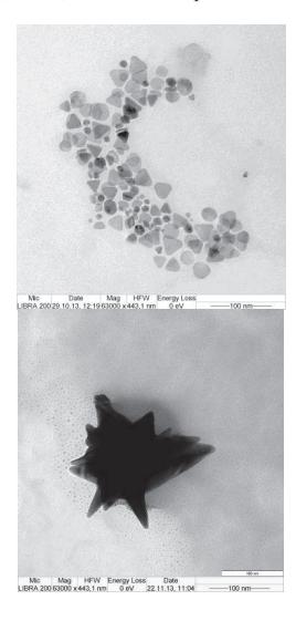
The colloidal solutions were prepared by adapting the synthesis procedures described in [1-2]. Silver nitrate or chlorauric acid was reduced to nanoparticles by a two-step procedure: synthesis of seed solution and growth of the particle. Shapes and sizes of the particles were analysed via transmission electron microscope LIBRA 200 from Carl Zeiss SMT.
Samples were measured with several spectrometers from Bruker (Germany): Tensor 27, Hyperion 3000 (IR microscope), IFS66, and a Raman spectrometer RamanRxn1TM from Kaiser Optical Systems Inc. (USA).