Gold nanoparticles are known for their unique photoluminescence, plasmonic properties [1], easy surface modification and functionalization with wide spectrum of organic molecules and drugs [2], low toxicity to organisms [3]. Due to these properties gold nanoparticles are very promising for cellular imaging, gene therapy, molecular diagnostics and therapy using targeted drug delivery [1]. However, mechanisms of accumulation and cytotoxicity of gold nanoparticles are not completely explored yet, therefore the purpose of this study was to synthesize MES (2-(N-morpholino) etanesulfonic acid) capped gold nanoparticles (Au-MES NP) and analyse their dynamics of accumulation in cancer cells.
Au-MES nanoparticles were synthesized using modified P.C. Chen et. al. synthesis protocol [4]. Spectral properties of Au-MES nanoparticles were analyzed: photoluminescence intensity of Au-MES nanoparticles diluted with cell growth medium was lower in comparison with PL intensity of Au-MES NP diluted with deionized water. This could be due to interaction of Au-MES NP with components of cell growth medium [5]. Accordingly, accumulation of Au-MES nanoparticles in cells can be affected by their interaction with compounds of cell culture medium.
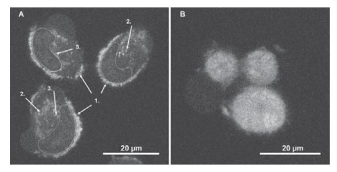
Investigating dynamics of Au-MES NP accumulation of MiaPaCa-2 cancer cells were incubated with Au-MES nanoparticles (volume ratio of cells growth medium to Au-MES nanoparticles solution was 3:1). Confocal fluorescence images (microscope Eclipse TE2000-S, $\lambda_{ex}$ = 404 nm) were taken at 3, 6, 9 and 24 hours after the incubation with gold nanoparticles. The adherence to the cell membrane (Fig. 1A, arrow no. 1); formation of granulated clusters (Fig. 1A, arrow no. 2) and localization of granulated clusters at the perinuclear region (Fig. 1A, arrow no. 3) after 3 hours of incubation of Au-MES nanoparticles were recorded. Distribution of Au-MES nanoparticles in cells after 3 hours correlates with stages of endocytosis mechanism. Diffuse distribution (including cell nuleus) with accumulation of Au-MES nanoparticles in several places near plasma membrane were visualized after 6 and 9 hours of incubation. After 24 hours of incubation homogeneous diffuse distribution of Au-MES NP in cancer cells was observed (Fig. 1B).