
The identification of pigments, ground layers and binders in polychromy of artworks is very important for restoration and conservation. Raman spectroscopy method is a powerful technique in analysis of artifacts and is used widely for more than 30 years [1]. Proton induced X-ray emission (PIXE) analysis is accurate method to investigate element analysis [2]. In this study the samples of Egyptian sarcophagus of the god's Amon singer (Thebes, XI - IX century BC) and 300 years old baptistery were investigated by Raman and PIXE spectroscopic methods (Fig. 1).
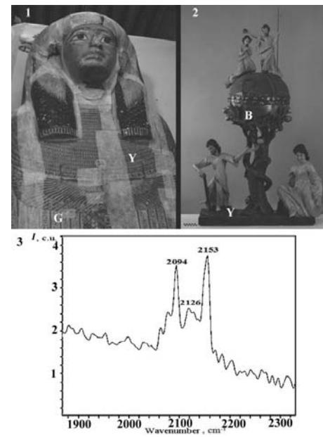
The results of sarcophagus green sample PIXE analysis showed that green sample consists of mostly of copper (37%) and calcium (23%). Investigation using Raman spectroscopy revealed that green pigment is a mixture of azurite (Cu3(CO3)2(OH)2) and malachite (Cu2(OH)2CO3). Raman spectra of yellow sample of sarcophagus showed that yellow pigment is an orpiment (As2S3) with impurities of goethite (Fe3+O(OH)).
Yellow and blue samples of baptistery were investigated using PIXE analysis. Results showed that yellow sample consists mostly of lead (64.1%), sulfur (17.2%) and calcium (13.9%). Blue sample mostly has calcium (87.4%) and iron (5.3%). Raman spectroscopy method showed that yellow pigment is lead white (Pb3(CO3)2(OH)2) and blue pigment is Prussian blue (Fe4[Fe(CN)6]3).
In conclusion, Raman spectroscopy and PIXE analysis combined together are effective methods for identification of pigments, ground layers and binders, but has some shortcomings in ways of sample preparation, which are under consideration in this work.