
Bioimpedance spectroscopy methods with using interdigital capacitive sensors are perspective for non-invasive analysis in biomedical applications, for example in pre-clinical drug development [1]. They can provide information on functional, electric and proliferative activity of cells [2]. Nowadays capacitive sensors are utilized in studying of toxicity of pharmaceutical drugs [3]. However, the using techniques are based on detection of cells quantity in a sample demand long periods. They are not allow to register the early functional changes arising in cells at their stimulation or disruption. Thus, the purpose of present work is to develop an express method for registration of functional and electric activity of cells using interdigital capacitive sensors.
The sensor's biosensitive dielectric nanostructurized coat containing cerium ions and multiwall carbon nanotubes modified by carboxyl groups was created by Langmuir-Blodgett technology. Cellular monolayers were formed on a sensor's surface at growing of C6 rat glioma cells. The registration of frequency and capacity characteristics of the sensor was carried out by means of the impedance analysis using electro-physical measuring device based on the pulse arranged RC-oscillator.
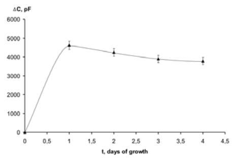
As shown in the Fig.1, addition of cell suspension in the medium leads to increment of sensor's capacity by increasing the concentration of ions (cells can be considered as macroions). The formation of a continuous cellular monolayer is observed on the second day of cell growth and is accompanied by electric-field shielding effect (EFSE), revealing as sensor capacity (C) decrease. The degree of EFSE is gradually rising on 3 and 4 days with increase of cellular monolayer density. However, the degree of EFSE depends on a content of medium used and increases in following sequence: C(0,15 mol/l NaCl)<C(0,15 mol/l NaCl with 2,5 mmol/l CaCl2) < C(Erla medium without 2,5 mmol/l CaCl2 (pH=7,4))<C(Erla medium)<C(minimal Egla medium). We proposed that such dependence is associated with changes in value of membrane potential, which characterizes the electric activity of cells.
The functional activity of cells can be estimated based on metabolic activity at their stimulation. In [4] it has been shown by means of chemiluminescent analysis that stimulation of glioma cells at menadione addition leads to production of superoxide anions in extracellular medium. We registered an increase of C at menadione action on glioma cells. General view of the kinetic dependence C(t) is consistent to the corresponding dependence obtained by the chemiluminesent analysis.
Thus, the developing bioimpedance spectroscopy method with using interdigital capacitive sensors allows to receive information on degree of cellular monolayer formation, metabolic and electric activity of cells.