
Urinary stone disease is common worldwide with prevalence of about 10 % of human population [1]. A key condition for urolithiasis to start is urine oversaturation with some specific chemical components, which leads to crystal growth and aggregation. Early discovery together with the identification of the exact chemical composition of urinary sediments could be crucial for taking appropriate preventive measures that inhibit kidney stone formation or growth processes. Optical microscopy, which is now the “gold standard" applied for the analysis, is unreliable as various crystals with the same chemical composition can have different physical appearance and vice versa.
The analysis of the recorded IR spectra allows obtaining objective information about chemical composition of the sediments even if they are multi-component. However, crystals of small size (10 – 100 μm) are strongly scattering the IR radiation. Therefore, the spectra are often distorted by scattering effects (peak shifts, first-derivative features of the bands and/or baseline oscillations) which can influence the results of the analysis by limiting complete characterization of the deposits, particularly, when they are multi-component. Such effects have been previously observed in the IR spectra of biological cells and human lymph node tissue [2, 3].
Transmission mode of the IR microscope Hyperion 3000 combined with IR spectrometer Vertex 70 was employed to record the IR absorption spectra of the urinary sediments. We analyzed urine samples of 136 healthy persons' urinary sediment. Urine samples were collected and were centrifuged leaving mostly urea and urine sediments in the samples. Then, after being placed on a filter (Whatman 542), the urine was left for 24 hours to dry. Then, the isolated crystals of the sediments were collected from the surface of the filter and transferred to the transparent for IR radiation plate (ZnSe) to analyze them.
We used the correction algorithm described in [3] for the correction of IR spectra of urinary sediments. The spectral corrections were performed using MATLAB package. Every IR spectrum measured was compared to the corresponding reference pure chemical compound spectrum (recorded using KBr pellet technique).
The results presented in figure 1 display an example of optical image of urinary deposit crystal constituted from brushite and corresponding IR absorbance spectrum obtained after crushing it between two ZnSe plates. Baseline oscillations and first-derivative effect can be observed in the spectrum which suggests the presence of the Mie scattering.
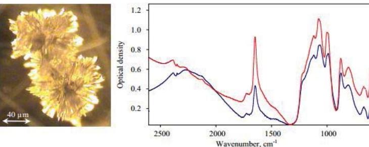
The spectrum obtained after the correction procedure is presented in figure 1. It can be seen that optical density the scattering effects are considerably diminished. Similar results were obtained by applying the correction procedure to the spectra of the sediments of various compositions. The analysis of the corrected spectra provides more reliable results and thorough characterization of the sediments can be performed.
In this work we have deduced that quality of infrared absorption spectra of urinary sediments can be improved when correction of the spectral baseline is performed by taking Mie scattering effects into account; thus, reliable chemical analysis can be carried out.