
Chemical imaging is an important technique in various fields of study. It provides information about chemical composition and structure of the analyzed sample. Infrared (IR) spectral imaging in specular reflection mode, however, is suffering from the fact that the spectral bands (so called Reststrahlen bands) in the specular reflection spectra resemble first derivative of the absorption spectrum and they are shifted towards lower wavenumbers [1]. Standard Kramers-Kronig (KK) procedure used to recreate absorption spectral bands from the Reststrahlen bands is not applicable to correct the raw spectra of urinary calculi as they contain diffuse reflection component arising from rough surface of the cross-section. In this work, urinary stones with various surface roughnesses were analyzed by infrared microspectroscopy and the bands in different spectral regions were used to create chemical images in order to determine the influence of the diffuse reflection to the imaging results. For study the influence of the diffuse reflection to the imaging results, the cross-sectioned stone was polished with sand paper of various roughnesses. The measurement with the optical profilometer allowed determining the degree of roughness after each polishing: P220 - 20 μm, P240 – 15 μm, P400 – 7-10 μm, polishing glass – 5-10 μm. The influence of the diffuse reflection to the reflection spectra can be estimated when the KK transform is applied. The amount of the diffuse reflection decreases when the ratio of the wavelength of the incident radiation with the size of the surface roughness increases. The results of the KK transform applied to each spectral band reveal that the longer wavelength of the spectral band is better correction of its shape after KK transform which in turn increases quality of chemical image of the surface.
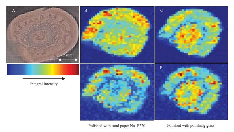
The chemical imaging results are presented in figure 1. The images, representing distribution of calcium oxalate, obtained for the stone with roughness size of 20 μm reveal clear difference (Fig. 1 B, D): when the spectral band corresponding to a longer wavelength is used to create the chemical image, the contrast is higher and the boundaries of calcium oxalate domains can be distinguished. When the surface roughness becomes smaller the chemical images obtained from different spectral bands are of similar quality (Fig. 1 C, E). It can also be seen in figure 1, that the smaller roughness of the surface, is reasoning higher resolution of the chemical images. In this work, we have demonstrated that the results of the chemical imaging depend significantly on the amount of the diffuse reflection contributing to the infrared reflection spectra. However, by choosing an appropriate spectral band at largest possible wavelength in comparison to the size of the roughness, reliable chemical imaging results can be obtained.