
The application of nanotechnology in medicine is a great interest of many researchers. One of the most promising research fields is application of magnetic nanoparticles (MNP) in early cancer diagnostics. Magnetic nanoparticles have unique magnetic properties, such as superparamagnetism, which depend on their size, geometry and chemical composition. Nowadays there are various disciplines, where magnetic nanoparticles could be used, but the main goal is to use magnetic nanoparticles as a contrast agent in oncology. The main issues, which have to be overcome, before using MNP in cancer diagnostics and therapy, are their toxicity, biocompatibility and chemical activity [1]. MNP do not fluorescent, so their imaging using optical microscopy methods, is complicated. In this study, few different imaging methods were applied to detect intracellular localization of superparamagnetic cobalt ferrite nanoparticles (SPIONs).
Firstly, SPIONs were incubated with different cell lines – liver carcinoma (MH-22A), ovarian cancer cells (A278) and fibroblasts (NIH3T3). Then two different imaging methods, such as differential interference contrast microscopy and staining MNP with Prussian blue (Fig.1, a, b), were investigated. Images were obtained using bright field function of confocal microscope.
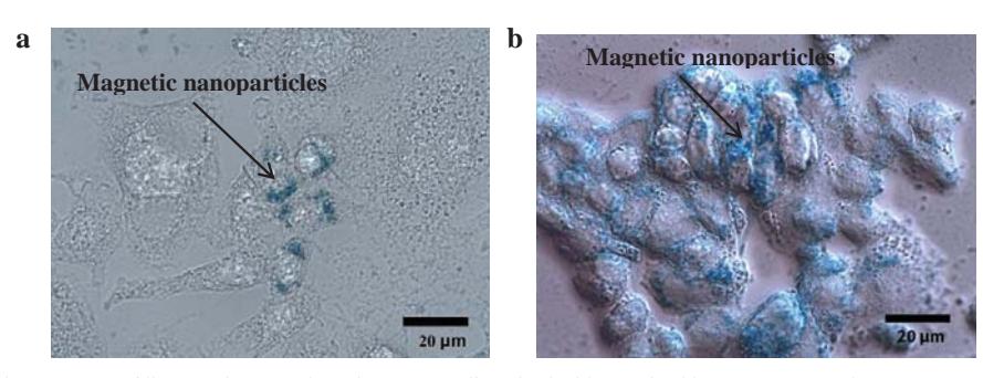
Images obtained using differential interference contrast microscopy show, that this method is not suitable for evaluation of intracellular localization of superparamagnetic cobalt ferrite. The main reason is that the light, which passes through cells, is equally absorbed and scattered by vesicles full-filled with MNP or nutrients, so in images intracellular SPIONs appear as the same black spots.
Intracellular labeling of SPIONs with Prussian blue dye showed different amount of accumulated MNP in normal and cancer cell lines. Fig 1 a, b show, that after 24h SPIONs tend to accumulate in the perinuclear region of – MH-22A and A278 cell lines. These results also revealed that MNP are more likely to accumulate in ovarian cancer cells, than in liver carcinoma cells, or fibroblasts. So it would be more efficient to use superparamagnetic cobalt ferrite nanoparticles with ovarian cancer cells, than with other ones.