
Quantum dots (QD) are photoluminescent semiconductor nanocrystals with specific physicochemical properties which depend on their spatial parameters and chemical composition and which make them advantageous fluorophores for many applications. QD are usually made of heavy metals core and stabilized with biocompatible ligands to reach biostability in biological medium. However under environmental conditions such as UV irradiation organic coatings may be destabilized and heavy metal ions may be released and induce toxicity for the biological systems. It was shown that healthy skin provides a barrier that protects from passage of QD to the organism but skin damages may increase the possibility of QD penetration into the deeper layers. Combined exposure of QD and UV irradiation may lead to the adverse physiological effects in two ways: it might destabilize QD and induce the penetration of the toxic metal ions through skin and/or it might induce dermal damages which decrease the functionality of the skin barrier.
In this study we present the photostability of two types of QD with different structure i.e., CdTe core QD covered with mercaptopropionic acid (CdTe-MPA) and CdSe/ZnS core/shell QD covered with amphiphilic polymer and polyethylene glycol layer (CdSe/ZnS-PEG). We also assessed the accumulation of QD in the skin tissues after topical application under UV irradiation.
Histological examination of UVB irradiated (260-320 nm) skin samples showed that UVB induced skin damage such as: disruption of sebaceous glands, appearance of apoptotic cells, pyknotic nuclei, clear cytoplasm, hyperemia [1]. Spectroscopic investigations revealed that during UVB irradiation photoluminescence (PL) intensity of CdTe-MPA solution increased, the full width at half-maximum of the PL spectrum expanded and the peak of the PL spectrum shifted to the short wave side (Fig. 1A) when compared with the dark conditions. Meanwhile in case of CdSe/ZnS-PEG PL intensity also increased during irradiation but there were no significant changes of spectral shape (Fig. 1B). It shows that nanocrystal structure of these QD was not affected.
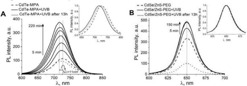
Spectroscopic changes of the CdTe-MPA QD show that UVB irradiation might damage the core. The degradation of the core is associated with the release of cytotoxic ions. [2] It raises the risk of potential harmful health effects if QD are applied in vivo. Differences in photostability between the two types of QD are related to the chemical composition: core/shell structure protects QD from oxidation and polymer ligands are resistant to UV damage.
Confocal fluorescence microscopy revealed that CdSe/ZnS-PEG QD distributed in the stratum corneum of the epidermis, in the superficial part of the hair channel and in the skin folds. However, UVB had no significant impact on QD localization in the skin when compared with the control non-irradiated skin samples.
We conclude that UVB irradiation causes temporary PL photoenhancement of CdTe-MPA and CdSe/ZnS-PEG QD. It is followed by shortwave shift of CdTe-MPA QD spectral band indicating the destabilization of the core which is related to the release of toxic heavy metal ions. It raises the risk of potential adverse health effects. Meanwhile CdSe/ZnS-PEG QD retain intact nanocrystal structure under UVB exposure. UVB irradiation does not alter QD localization in mice skin in vivo despite of physiological dermal damage.