
Plasmon-enhanced luminescence of molecular probes in plasmonic nanostructures is a highly developing field providing a number of new methods in chemical and biomedical analysis. The proximity of a metal nanobody gives rise to an increase in local electromagnetic radiation intensity, changes the probability of spontaneous photon emission, and also promotes multiple enhancement of nonradiative relaxation of an excited state [1]. All three effects result from local modification of the dielectric function of space upon insertion of a metal nanobody which is capable to support plasmon oscillation. Theoretical model of luminescence enhancement/quenching for luminophores near metal spherical nanoparticles stated that there is a strong correlation between intensity of fluorescence and silver particle size as well as distance "fluorophore – metal" [2].
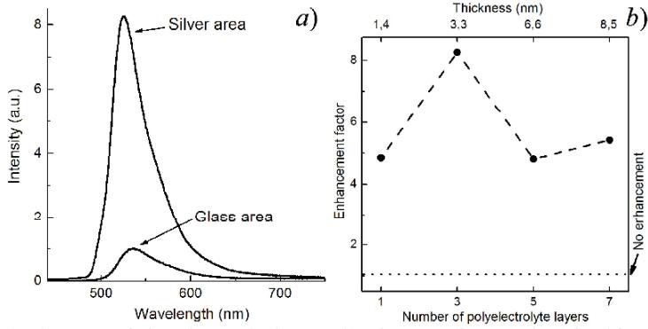
In this paper we investigated plasmon-enhanced luminescence of Alexa Fluor 488 molecules that are widely used in biomedical applications. For this purpose multilayer nanostructures “silver nanoparticles – polyelectrolytes – Alexa Fluor 488 molecules" were prepared. The distance between molecules and metal nanoparticles was controlled by varying number of counter-charged polyelectrolyte layers. Average diameter of the particles was about 40 nm (the optical density spectrum features the maximum near 415 nm) according to scanning electron microscopy data. The dielectric spacer was developed only up to 1, 3, 5 and 7 polyelectrolyte layers because of the diminution of the plasmonic effects with distance increasing. It was found that all fabricated and examined silver containing samples enhanced Alexa Fluor 488 luminescence in comparison to the reference samples with the same polyelectrolytes layers on glass substrates without silver coatings. Photoluminescence intensity of Alexa Fluor 488 molecules as a function of the number of polyelectrolyte layers is shown in Figure 1b. 8.2-fold maximal enhancement was observed for the sample containing three polyelectrolyte layers (corresponding thickness is 3.3 nm) (Figure 1a).