
Medical imaging techniques have great importance to the course of the cancer treatment process. One of the imaging methods is Nuclear Medicine, where radioactive materials such as Technetium-99m (99mTc) are used. However, this method has low resolution and the image can be recorded only on the screen.
In cases where the boundaries of the object have to be determined more precisely, fluorescing materials can be used, for their better resolution and ability to directly visualize of the object [1]. Fluorescent nanoparticles (NP) are of great scientific interest at the moment, for they can be bound to biomolecular structures and delivered to the selected targets. Radioactive metals such as 99mTc can be used for labeling these NPs allowing to apply dual imaging modalities. This combination would give the opportunity to use dual imaging of structures and help surgeons visualize the tumor.
In this research dual imaging NPs were synthesized using photoluminescent gold bovine serum albumin nanoclusters (Au-BSA NC) and “cold” Technetium-99 (99Tc). Au-BSA nanoclusters were synthesized using tetrachlorauric acid (HauCl4), BSA and sodium hydroxide (NaOH) as a reducing agent [2]. Synthesis of Au-BSA-99Tc NP was carried out using 1 ml of 99Tc, 1 ml of Au-BSA ($3.6\times10^{-7}$ mol/ml), 0.5 ml SnCl2 (0.4 mg/ml); Saline (0.9% NaCl). The content of the SnCl2 in Au-BSA-99Tc solution varied. To make stannous solution, 4 mg of SnCl2 were diluted with 10 ml of Saline. Synthesis was done by mixing Au-BSA with the stannous solution and then adding 99Tc [2].
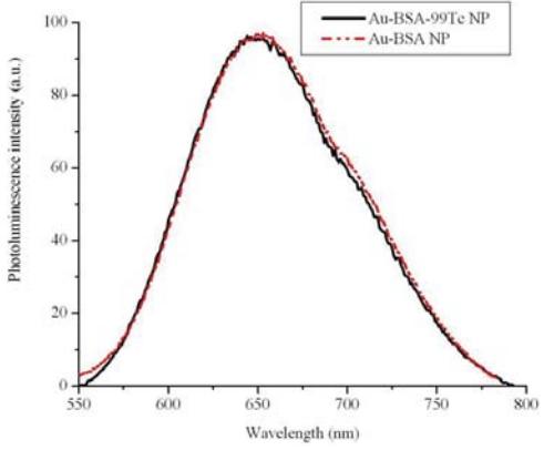
To evaluate the photoluminescence properties of synthesized Au-BSA-99Tc compound spectrophotometric analysis were performed.