
Raman spectroscopy is a commonly used tool for determining the composition of various substances. Unfortunately, the measurements are only effective if the molar concentration of the substance is high enough. Typically, the detection limit is $1 \div 10$ mM. However, the signal of the measurements can be significantly increased by applying the surface enhanced Raman scattering (SERS). In this work, a couple of chemical constituents of urinary sediments: L-Cystine, Urea, were analysed by this method.
Elevated concentrations of urinary sediments indicate a higher risk of formation of renal stones [1]. Identification of sediments can be performed by analysis of their constituents. As the concentration of these constituents is far too small for the conventional Raman spectroscopy, SERS method had to be used to measure their spectra. Two samples of $10^{-4}$ M and one sample of $10^{-3}$ M were studied.
Surface enhanced Raman spectroscopy can be applied by using the nanoparticle colloids of gold (Au), silver (Ag) or copper (Cu). In this work silver nanoparticle colloid, prepared by Lee and Meisel method [2], was used. To intensify the aggregation of silver nanoparticles the 0.05 M of sodium chloride (NaCl) was added to the solution of colloid and sample. The spectra of dried samples were measured with Bruker FT-Raman spectrometer “Multiram” and 1064 nm laser was used for the excitation. Conventional Raman and SERS spectra of urea is presented in Fig. 1.
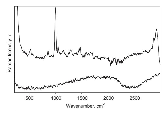
Upon processing the registered spectra it was found that the enhancement factor for urea samples varied from a hundred to a thousand times. This variability of enhancement factor can be explained by the different sizes of the silver nanoparticle aggregates formed in different experiments. This finding calls for further experiments and optimization of the experimental parameters. New SERS experiments with electrochemical cell are intended in the near future.