
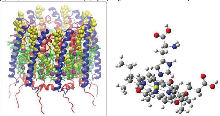
Bacteriochlorophyll-a molecules are part of the LH2 complex. LH2 complex acts as an antenna that absorbs solar energy which is subsequently transferred to the reaction center. LH2 complex is composed of two kinds of polypeptides that form a cylinder connecting the edges of the membrane. Within this cylinder two rings of bacteriochlorophyll molecules can be found. Bacteriochlorophyll molecules are in close proximity with histidine groups from both chains of polypeptides, which affects bacteriochlorophyll group magnesium atom. Entire LH2 complex is shown in Figure 1 [1].
Histidine is one of the 20 neutral amino acids [2]. Imidazole ring of histidine chain is chemically unstable. It may have different tautomers with regard to its protonation and act as a proton donor, acceptor or proton-transfer agent. There are four different imidazole ring protonation forms (i.e. formally anionic imidazolate form).
This work is based on the interaction between bacteriochlorophyll-a and neutral histidine molecules from the LH2 complex. The structure used was taken from the LH2 complex in Protein Data Bank [3]. Bacteriochlorophyll-a (structure called bcl1601) and histidine ($\beta$-his30) are shown in Fig. 2. For visualization, the GaussView program was used [4].
The main goal of this study is the investigation of the influence of histidine molecules to bacteriochlorophyll spectrum. The distance between protein chains bearing histidine and bacteriochlorophyll were fixed at different distances and the geometry of remaining dimer structure was optimized. For this purpose, several methods have been used: BOP TDDFT with LC correction, TDDFT B3LYP, and FMO [5]. Gaussian and GAMESS quantum chemistry packages [6,7] of Vilnius University Physics faculty supercomputer [8] were used in this work.
It was determined that the TDDFT BOP/6-311 method with LC correction gives the electronic excitation energies closest to experimental results. Five different structures were selected by varying the distance between protein chains. It was determined that the energies and oscillator strength of the two lowest electronic excitations practically do not change during the investigated deformations. The distance between the magnesium atom of bacteriochlorophyll and the nitrogen atom of histidine in the most stable histidine-bacteriochlorophyll dimer is 2.19 Å.
The work is still expected to continue by changing the histidine tautomers and by using the FMO method in order to take into account the influence of the surrounding protein environment to BChl.